
FIGURE 6
- ID
- ZDB-IMAGE-210103-17
- Publication
- Narumi et al., 2020 - Chemical-Induced Cleft Palate Is Caused and Rescued by Pharmacological Modulation of the Canonical Wnt Signaling Pathway in a Zebrafish Model
- All Figures
- Figures for Narumi et al., 2020
|
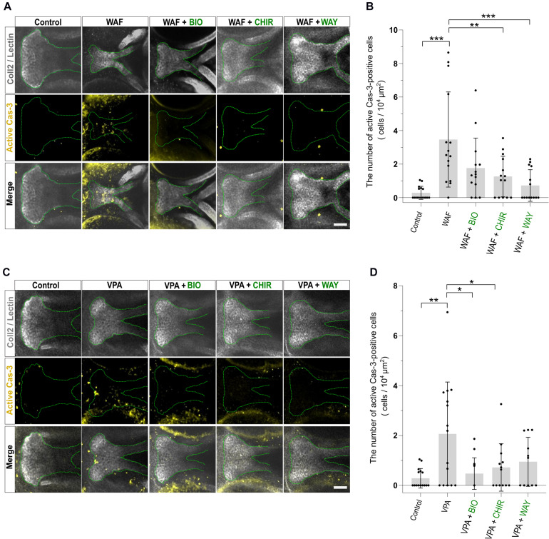
FIGURE 6
Apoptosis level was restored by combinatorial treatment with teratogen and Wnt agonists.