
FIGURE 2
- ID
- ZDB-IMAGE-210103-13
- Genes
- Publication
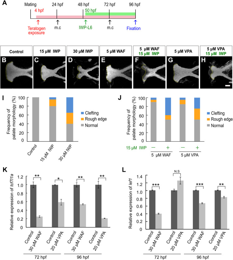
- Narumi et al., 2020 - Chemical-Induced Cleft Palate Is Caused and Rescued by Pharmacological Modulation of the Canonical Wnt Signaling Pathway in a Zebrafish Model
- All Figures
- Figures for Narumi et al., 2020
|
FIGURE 2
Chemical-induced cleft palate was induced by inhibition of the canonical Wnt signaling pathway.