IMAGE

FIGURE 8

ID
ZDB-IMAGE-201230-10
Source
Figures for Delbaere et al., 2020
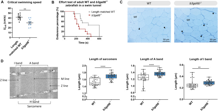
Image
Figure Caption

FIGURE 8

Functional and structural analysis of adult WT and b3galt6–/– zebrafish muscle. (A) The critical swimming speed and (B) the endurance performance was determined for b3galt6–/– and length-matched WT zebrafish. (C) Semi-thin cross sections, stained with toluidine blue, demonstrate the presence of a thicker endomysium (arrowhead), located between the muscle fibers (Mf) at the horizontal myoseptum of b3galt6–/– zebrafish at the age of 5 months. (D) Transmission electron microscope structure of a sarcomere with indications of the Z and M line and A, I, and H band. The sarcomere length was determined at 5 different loci per semi-thin section, for 6 semi-thin sections origination from 3 different zebrafish per genotype (N = 30). A significant increase in sarcomere length for b3galt6–/– is noted. Furthermore, both the A and I band show an increase in length for the mutant zebrafish. Data are expressed as box plots with min-to-max whiskers on which each individual data point is plotted. A Mann-Whitney U test was used to determine significance. ****P < 0.0001, **P < 0.01.

Figure Data
Acknowledgments
This image is the copyrighted work of the attributed author or publisher, and ZFIN has permission only to display this image to its users. Additional permissions should be obtained from the applicable author or publisher of the image. Full text @ Front Cell Dev Biol