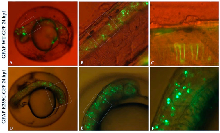
IMAGE

Figure 1

ID
ZDB-IMAGE-201229-79
Source
Figures for Candiani et al., 2020
Image
Figure Caption

Figure 1

GFP expression in transgenic glial fibrillary acid protein (GFAP) wildtype (WT) and GFAP R239C embryos. (A,B) The expression of GFAP WT-GFP protein is visible along the neural tube, with very few GFAP aggregates observed. (C) High magnification of embryo in (B) shows GFAP WT protein as filamentous structures. (DF) Several aggregates of p.R239C in the brain and along the neural tube are found. Magnification: 4× (A,D); 10× (B,E); 20× (C,F). High magnification are shown by rectangular shape.

Acknowledgments
This image is the copyrighted work of the attributed author or publisher, and ZFIN has permission only to display this image to its users. Additional permissions should be obtained from the applicable author or publisher of the image. Full text @ Genes (Basel)