IMAGE

Fig. 6

ID
ZDB-IMAGE-201212-11
Source
Figures for Domenichini et al., 2020
Image
Figure Caption

Fig. 6

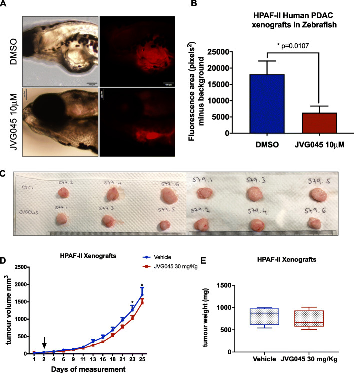
Effect of JVG045 on zebrafish and mouse xenografts.a Brightfield images and images showing DiL-labelled HPAF-II tumour xenografts in 5 days post-fertilisation zebrafish embryos untreated (DMSO) or treated with JVG045 (10 μM) for 3 days after injection of HPAF-II human pancreatic cancer cells. Scale bar 100 μm. b Graph showing the effect of 10 μM JVG045 in reducing the tumour burden in zebrafish embryos (n = 12), compared to control embryos (DMSO, n = 6). c Representative images of HPAF-II tumours from mouse xenografts receiving control treatment (vehicle) or treated with JVG045 (30mh/kg) for 25 days. d Graph showing tumour growth of HPAF-II xenograft mouse models treated with vehicle (n = 8) and 30 mg/kg JVG045 (n = 9). e Graph showing the tumour weight at the end of the experiment of HPAF-II xenograft mouse models treated with vehicle (n = 8) and 30 mg/kg JVG045 (n = 9). Results are expressed as Mean ± SEM

Acknowledgments
This image is the copyrighted work of the attributed author or publisher, and ZFIN has permission only to display this image to its users. Additional permissions should be obtained from the applicable author or publisher of the image. Full text @ J. Exp. Clin. Cancer Res.