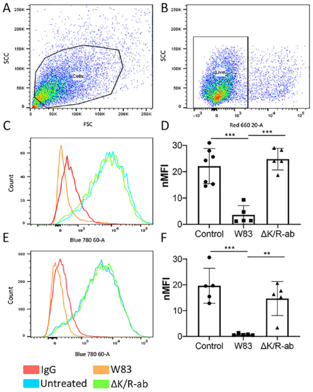
IMAGE

Figure 4.

ID
ZDB-IMAGE-201208-49
Source
Figures for Farrugia et al., 2020
Image
Figure Caption

Figure 4.

Loss of endothelial cell surface PECAM-1 by W83 whole bacteria and outer membrane vesicles (OMVs) is mediated by gingipains. Following a 1.5-h exposure to W83 or ΔK/R-ab whole bacteria or OMVs, human microvascular endothelial cells (HMEC-1) were removed from tissue culture plates and subjected to flow cytometric analysis for PECAM-1 cell surface abundance. Cells were gated using (A) side-scatter (SSC) and forward-scatter (FSC) voltages, then for (B) cell viability using TO-PRO-3 live/dead staining. Untreated cells were used as controls. Representative histograms and bar chart of 10,000 gated cells showing that PECAM-1 cell surface abundance is significantly decreased upon treatment with (C, D) W83 whole bacteria and (E, F) W83-derived OMVs compared to ΔK/R-ab-treated equivalents and untreated controls. PECAM-1 cell surface abundance was similar on HMEC-1 treated with ΔK/R-ab whole bacteria or ΔK/R-ab-derived OMVs to those observed on untreated controls. Data in D and F are presented as mean ± SD normalized median fluorescence index (nMFI) from 5 independent experiments with statistical significance determined by a 1-way analysis of variance with Tukey’s post hoc multiple comparisons test. **P < 0.01. ***P < 0.001.

Acknowledgments
This image is the copyrighted work of the attributed author or publisher, and ZFIN has permission only to display this image to its users. Additional permissions should be obtained from the applicable author or publisher of the image. Full text @ J. Dent. Res.