IMAGE

FIGURE 3

ID
ZDB-IMAGE-201121-43
Source
Figures for Kuroda et al., 2020
Image
Figure Caption

FIGURE 3

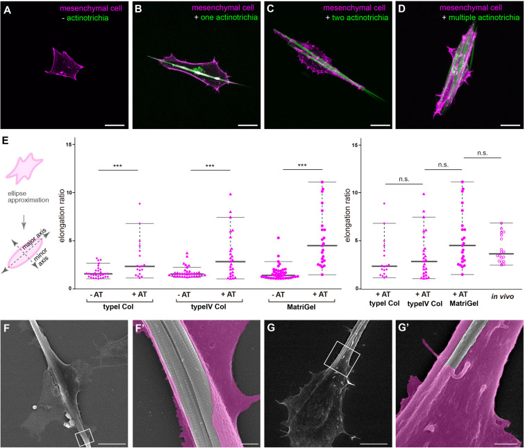
Interaction between mesenchymal cells and actinotrichia in vitro.(A–D) The morphology of primary cultured mesenchymal cells at day 2 after culture on a Matrigel-coated dish. Mesenchymal cells and actinotrichia were isolated from TG larval fins [TG; 5xand1(MC]: Lifeact-mCherry/5xand1(2P): And1full-GFP). (A) In the condition without actinotrichia, mesenchymal cells showed the symmetric morphology. (B–D) In the condition with actinotrichia, mesenchymal cells elongated along a single actinotrichia (B), two actinotrichia (C) and multiple actinotrichia (D). (E) Cell aspect ratio of mesenchymal cells. Mesenchymal cells in contact with actinotrichia had a more elongated shape compared to the cells not in contact with actinotrichia. (F–G′) SEM images of cultured mesenchymal cells holding the actinotrichia on a Matrigel-coated dish. The actinotrichia was surrounded with the plasma membrane of the mesenchymal cell (F) and partially embedded inside the mesenchymal cell (G). The magnified images in white box are shown. P-values: ***P < 0.0001. AT, actinotrichia. Scale bars: 20 μm in (A–D), 10 μm in (F,G), 1 μm in (F′,G′).

Acknowledgments
This image is the copyrighted work of the attributed author or publisher, and ZFIN has permission only to display this image to its users. Additional permissions should be obtained from the applicable author or publisher of the image. Full text @ Front Cell Dev Biol