IMAGE

Figure 7

ID
ZDB-IMAGE-201121-137
Source
Figures for Abdul Satar et al., 2020
Image
Figure Caption

Figure 7

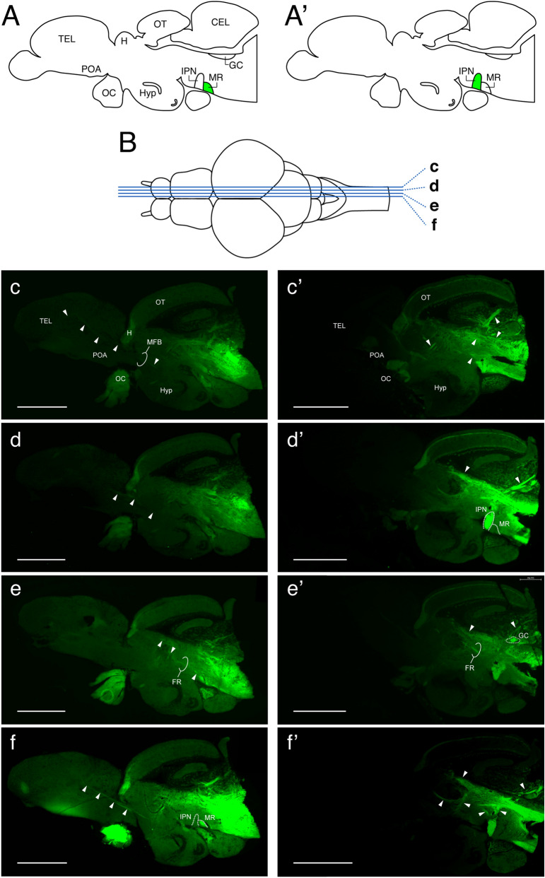
Tracer-labelled neuronal projection from the median raphe. (A,A’) Schematic sagittal view of the zebrafish brain illustrating the application sites of the neural tracer: the median raphe (MR, A) and interpeduncular nucleus (IPN, A’). (B) A schematic horizontal view of the zebrafish brain illustrating levels at which sagittal sections (cf and c’f’) were taken. (cf) Photomicrograph of mid-sagittal visualization of tracer (NeuroVue Maroon, green)-labelled neural fibers/tracks (arrow heads) from the MR. Tracer-labelled fibers/tracks were seen in the mesencephalon including the fasciculus retroflexus (FR, e), and in the rhombencephalon (ef). Tracer-labelled tracks were seen from the diencephalon towards the medio-dorsal region of the telencephalon which is the medial forebrain bundle (MFB, e,f). In addition, some fibers were also seen in the cerebellum (e,f). (c’f’) In the brain where the tracer was applied to the IPN (e’f’), tracer-labelled fibers/tracks were mainly seen in the dorsal region of the mesencephalon including the FR, and in the rhombencephalon and medulla spinal regions. In the rhombencephalon, fibers were seen in the griseum centrale (GC, e’). In addition, thick fibers were also seen in the cerebellum (c’f’). Scale bars 500 μm.

Acknowledgments
This image is the copyrighted work of the attributed author or publisher, and ZFIN has permission only to display this image to its users. Additional permissions should be obtained from the applicable author or publisher of the image. Full text @ Sci. Rep.