IMAGE

Figure 6

ID
ZDB-IMAGE-201121-136
Source
Figures for Abdul Satar et al., 2020
Image
Figure Caption

Figure 6

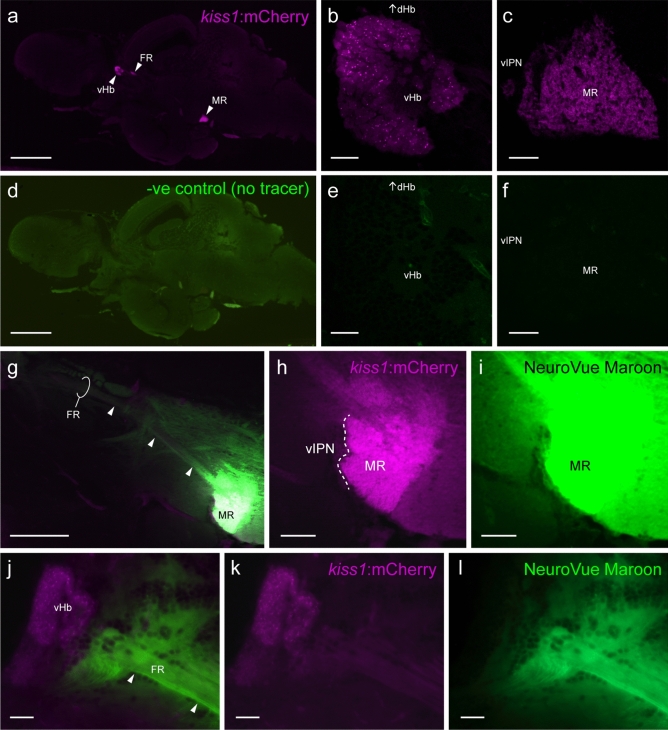
Neural tracer application into the median raphe. (af) Mid-sagittal visualization of mCherry (magenta)-labelled Kiss1 efferent projection from the ventral habenula (vHb, b) through the fasciculus retroflexus (FR) towards the median raphe (MR, c) in the brain of Tg(kiss1:mCherry) zebrafish without neural tracer implantation (df). (gi) A thin-strip of the nylon membrane-coated with NeuroVue tracer (NeuroVue Maroon, green) was selectively inserted into the MR (h,i). (jl) Photomicrograph of mid-sagittal visualization of tracer-labelled neural tracks (arrow heads) from the MR (g) towards the vHb (j). Scale bars (a,d) 500 μm; (b,c,e,f,jl) 20 μm; (g) 200 μm; (h,i) 50 μm.

Acknowledgments
This image is the copyrighted work of the attributed author or publisher, and ZFIN has permission only to display this image to its users. Additional permissions should be obtained from the applicable author or publisher of the image. Full text @ Sci. Rep.