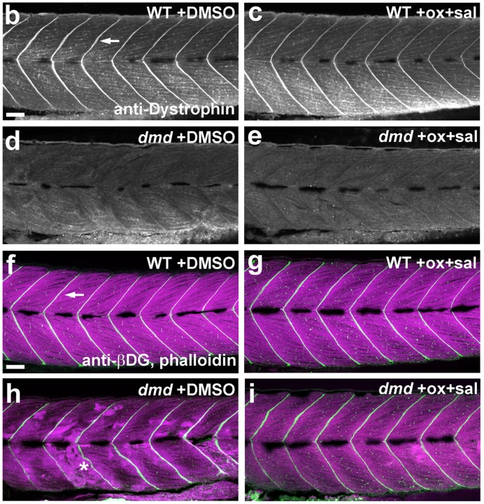
Fig. 7-2 Oxamflatin and salermide have dose-dependent effects, independent of dystrophin expression. a Graph of average normalized pixel intensities for treatments of dmd mutants with doses of oxamflatin and salermide. Control treatment is 1% DMSO. Chemicals were used between 0.5 μM and 4 μM, over two separate experiments. For each treatment condition, n = 4 replicates, with 2-11 dmd−/− embryos in each replicate. Plot shows the average normalized pixel intensity for each of the 4 replicate pools for each treatment. The vertical line separates the treatment conditions from the two experiments, each of which has its own WT + DMSO and dmd + DMSO controls. The dashed lines represent the average normalized pixel intensity for all of the DMSO-treated dmd animals (n = 26 and n = 30). Error bars represent standard error. Significance was determined using a one-way ANOVA test comparing each treatment group to the dmd DMSO control group with Dunnett’s correction for multiple comparisons. *p ≤ 0.029, **p = 0.0029, ***p = 0.0007 compared to dmd DMSO control. b-e Confocal images of anti-dystrophin staining in the trunk musculature of 4 dpf b WT + DMSO, c WT + oxamflatin and salermide, d dmd + DMSO, and e dmd + oxamflatin and salermide larvae. Lateral views, anterior to the left. Arrow points to dystrophin expression in the vertical myoseptum. All dmd+/+ animals showed normal dystrophin expression (WT + DMSO, n = 16; WT + ox+sal, n = 14) and all dmd−/− animals lacked detectable dystrophin expression (dmd−/− + DMSO, n = 23; dmd−/− + ox+sal, n = 18). Scale bar = 50 μm. f-i Confocal images of anti-β-dystroglycan (βDG) and phalloidin staining in the trunk musculature of 4 dpf f WT + DMSO, g WT + oxamflatin and salermide, h dmd + DMSO, i dmd + oxamflatin and salermide. Lateral views, anterior to the left. Arrow points to βDG expression (white) in the vertical myoseptum. Phalloidin staining of filamentous actin (magenta) shows the disrupted muscle structure in dmd mutants (* in h). All wild type animals (+/+ and +/−) showed normal β-dystroglycan expression (WT + DMSO, n = 27; WT + ox+sal, n = 26), and dmd−/− animals showed largely maintained β-dystroglycan expression (dmd−/− + DMSO, n = 9; dmd−/− + ox+sal, n = 14). Scale bar = 50 μm
Image
Figure Caption
Acknowledgments
This image is the copyrighted work of the attributed author or publisher, and
ZFIN has permission only to display this image to its users.
Additional permissions should be obtained from the applicable author or publisher of the image.
Full text @ Skelet Muscle

