IMAGE

Figure 5

ID
ZDB-IMAGE-201102-16
Source
Figures for Santhanam et al., 2020
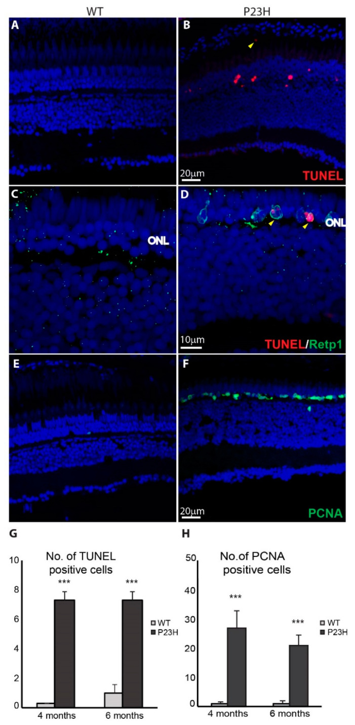
Image
Figure Caption

Figure 5

Cell death and cell proliferation in the P23H transgenic zebrafish. (A,B) Cell death detection using TUNEL staining shows TUNEL-positive dying cells (red) in the P23H zebrafish retina (4 months old). The yellow arrowhead shows a TUNEL-positive cell in the sub-retinal space. (C,D) Yellow arrowheads show the colocalization of Retp1 (green) with TUNEL labeling in the P23H transgenic fish. (E,F) PCNA immunostaining shows many PCNA-positive proliferating cells (green) in the ONL of the P23H zebrafish, but very few in WT. ONL: outer nuclear layer. (G) Numbers of TUNEL positive cells and (H) PCNA-positive cells per 210 µm image field in 4-month old and 6-month old WT and P23H retina (n = 6 fish per genotype; error bars are ± SD; *** p < 0.001).

Figure Data
Acknowledgments
This image is the copyrighted work of the attributed author or publisher, and ZFIN has permission only to display this image to its users. Additional permissions should be obtained from the applicable author or publisher of the image. Full text @ Cells