
Fig. 1
- ID
- ZDB-IMAGE-201029-1
- Publication
- Bagnat et al., 2020 - Development of a straight vertebrate body axis
- All Figures
- Figures for Bagnat et al., 2020
|
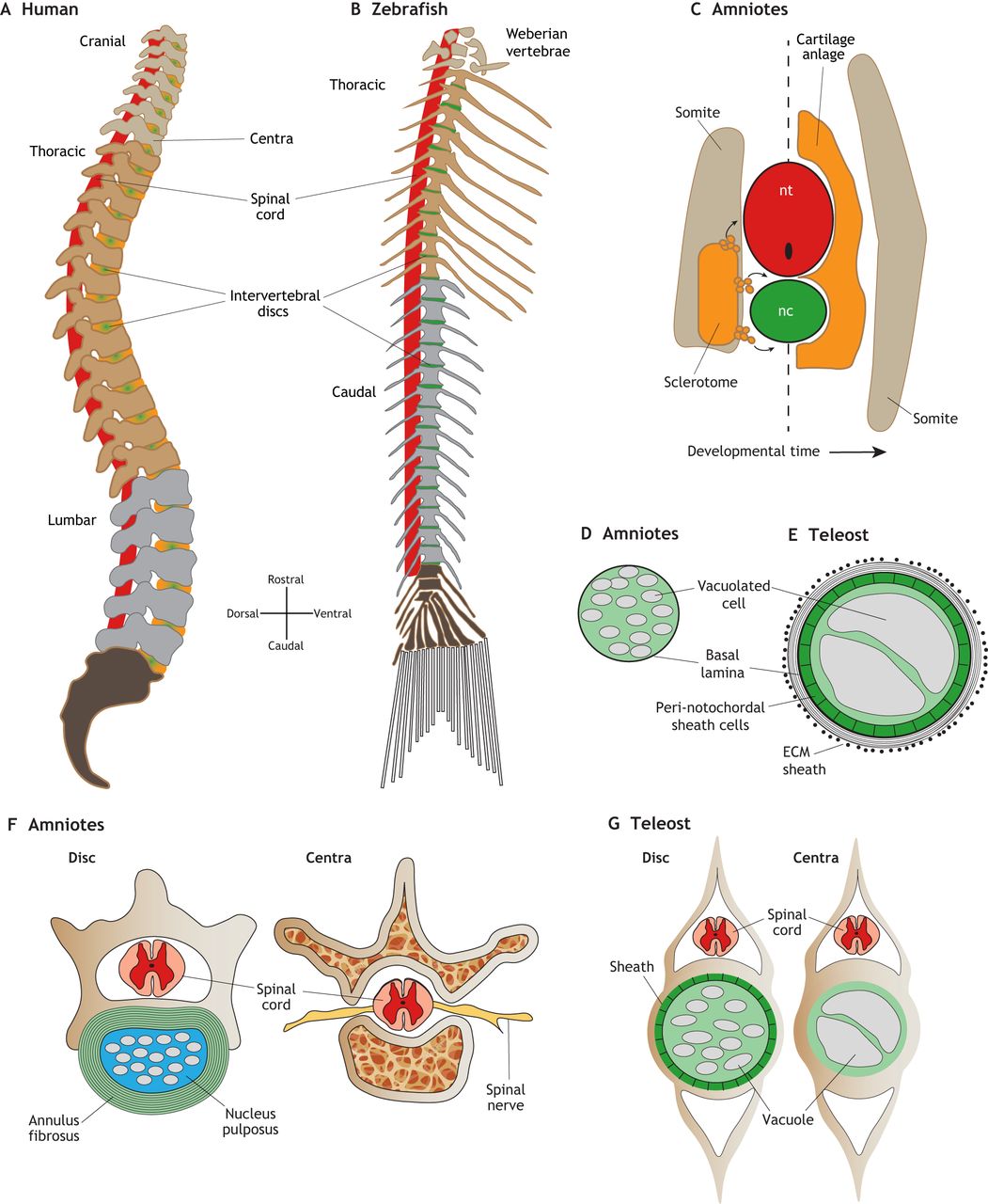
Fig. 1 Development and morphology of the spine in amniotes and zebrafish. (A) Lateral view schematic of a human adult spine. The intervertebral discs (green/orange) are largely derived from the cartilaginous anlage and notochord in mammals. (B) Lateral view schematic of a zebrafish adult spine. The intervertebral discs (green) are largely derived from the notochord in zebrafish. (C) Transverse view schematic of re-segmentation of the somite-derived sclerotome (orange) tissue which migrates out from the somite (left side of dashed line) to form the cartilage anlage, which wraps the spinal cord and notochord (right side of dashed line). The myotome region of the somite give rise to the axial muscles (tan). (D) Schematic of an amniote notochord in cross section, showing vacuolated cells and a basal lamina sheath. (E) Schematic of a teleost notochord in cross section, showing a larger notochord with large vacuolated cells enclosed by a peri-notochordal sheath cell epithelium, and a lamellar ECM sheath. (F) Cross-sections of mouse spine highlighting the multi-lamellar annulus fibrosus fibrocartilage layer (green), surrounding the nucleus pulposus (light blue), which contains notochord-derived cell lineages, the spinal cord (pink/red) and spinal nerves (yellow), and the trabecular bony regions. (G) Cross-sections of zebrafish spine showing the intervertebral disc (Disc) region, highlighting the sheath and small fragmented vacuolated cells that form the zebrafish disc and the large vacuolated cells within the vertebral body region.