
|
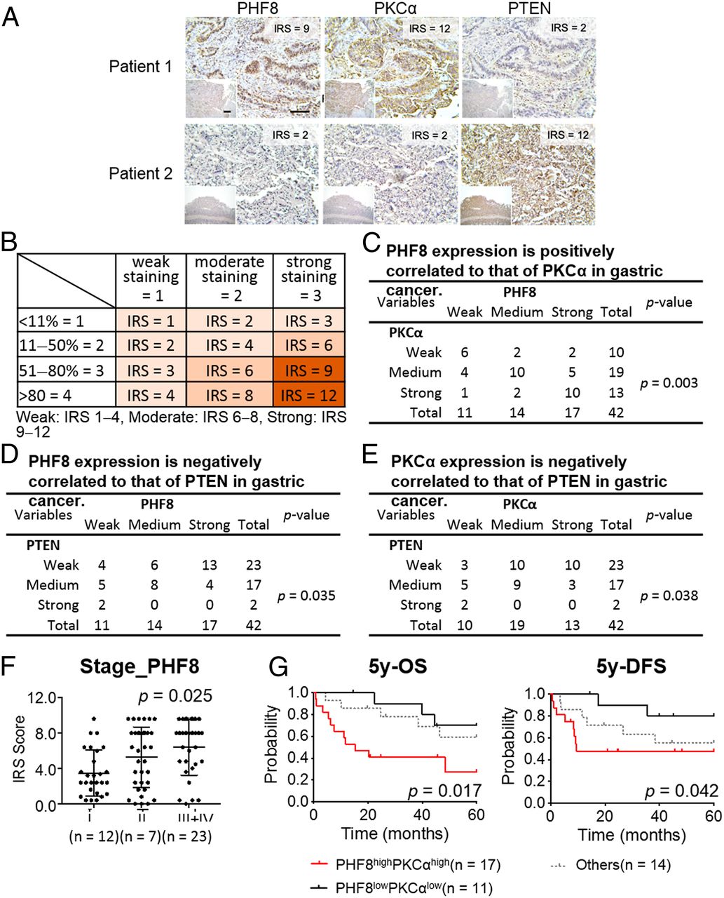
Fig. 7 Clinical relevance of PHF8, PKCα, and PTEN in 42 GC subjects obtained from CGMH. (A) Representative image of immunohistochemical profiles. PHF8, PKCα, and PTEN were immunostained for each of gastric tissue specimens (n = 42). (Scale bar, 100 μm.) (B) IRS score of GC samples. (C−E) The correlation of IHC signals for two-group comparisons: PHF8 and PKCα (C), PHF8 and PTEN (D), PKCα and PTEN (E). Statistical significance was evaluated using the χ2 test. (F) PHF8 expression is significantly correlated with tumor stage in patients with GC. Statistical calculation is conducted using one-way ANOVA analysis. (G) Five-year OS and 5-y DFS analysis according to the level of PHF8 and PKCα expression in patients with GC (n = 42). High, IRS ≥ 8; low, IRS ≤ 6. Statistical significance (PHF8highPKCαhigh vs. PHF8lowPKCαlow) was determined by log-rank test.