
Figure 3—figure supplement 2.
- ID
- ZDB-IMAGE-201021-11
- Publication
- Helker et al., 2020 - Apelin signaling drives vascular endothelial cells towards a pro-angiogenic state
- All Figures
- Figures for Helker et al., 2020
|
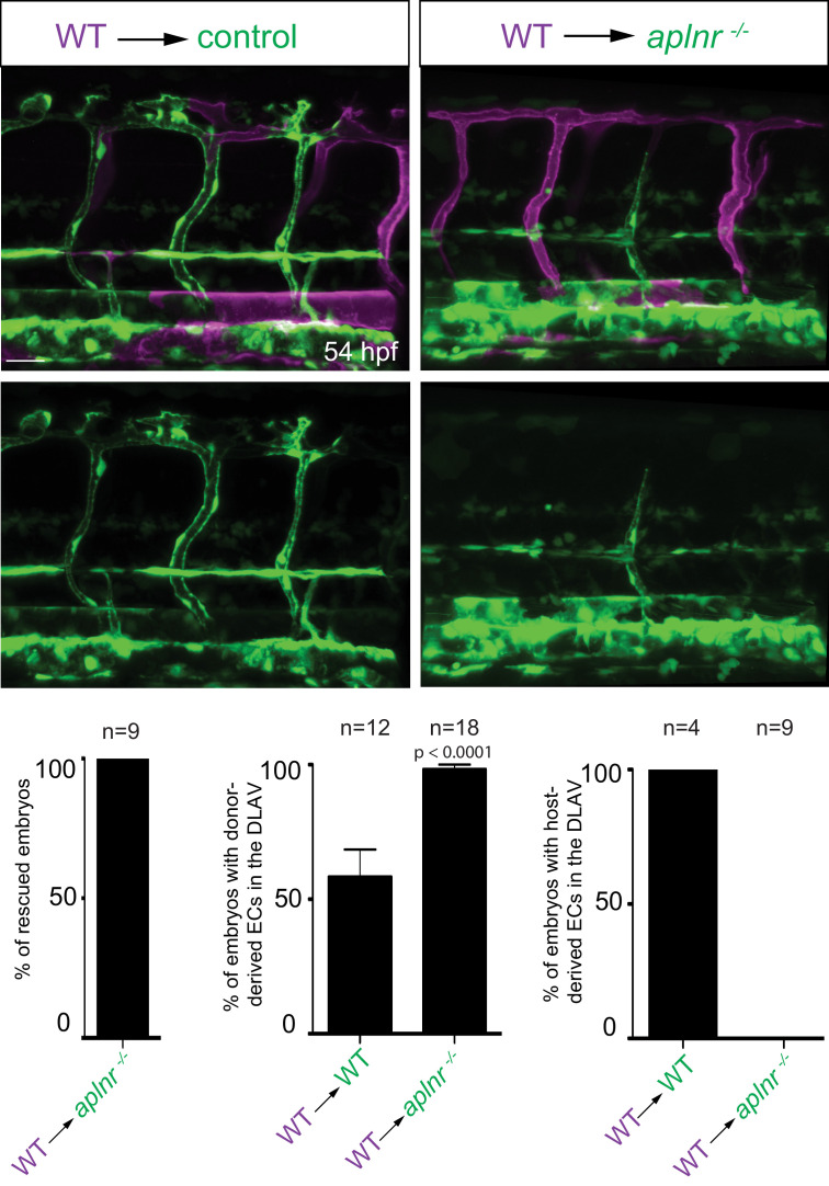
Figure 3—figure supplement 2.
Confocal projection images of the trunk region of Tg(fli1a:EGFP) embryos at 54 hpf. At the blastula stage, cells from Tg(kdrl:HsHRAS-mCherry) embryos were transplanted into host embryos obtained from Tg(fli1a:EGFP) aplnra +/-; aplnrb +/- incrosses. At 54 hpf, the mosaic embryos were imaged and the donor-derived ECs scored for their position. Notably, wild-type ECs transplanted into aplnr deficient embryos completely substituted for the lack of cells in the dorsal part of the vasculature at 54 hpf (Figure 3—figure supplement 1). Scale bar: 30 µm.
Apelin signaling functions cell-autonomously in ECs.