IMAGE

Fig. 1

ID
ZDB-IMAGE-201003-199
Source
Figures for Iwanami et al., 2020
Image
Figure Caption

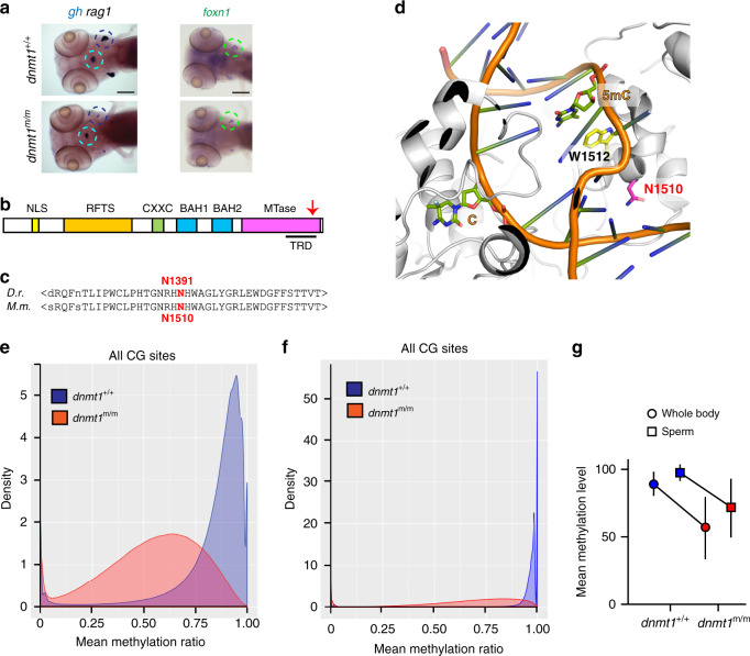
Fig. 1 Characterization of a hypomorphic allele of <italic>dnmt1</italic>.

a Impaired larval T cell development in mutant fish. Diagnostic whole-mount RNA in situ hybridization pattern 23in wild-type and dnmt1 mutant fish at 5 dpf using rag1 (thymus encircled in black), and gh (hypophysis encircled in blue) specific probes (left panel); hybridization pattern for foxn1, a marker of thymic epithelium59,60 (right panel). Scale bar, 100 μm. Panels are representative of 25 animals each. b Schematic of functional domains in the dnmt1 protein (not to scale); NLS, nuclear localization signal; RFTS, replication foci targeting site; CXXC, cysteine-rich domain; BAH, bromo-adjacent homology domains 1 and 2; MTase, catalytic domain. Arrow, approximate position of the amino acid replacement in the target recognition domain, TRD. c The asparagine residue mutated in the dnmt1t25501 allele occurs in an evolutionarily conserved region of the enzyme. d In the structure of the mouse Dnmt1 protein in complex with a hemi-methylated substrate, close apposition of the mutated asparagine residue to the substrate DNA in the catalytic site is observed26 (PDB ID: 4DA4). e, f Hypomethylation of cytosine residues in CpG dinucleotides of DNA extracted from whole body of 18 dpf larvae (e) (n = 3) and sperm of adult homozygous mutants (f) (n = 3). In (e) and (f), density refers to the fraction of CG sites with a particular methylation ratio. g Mean methylation levels of CG dinucleotides for DNAs shown in (e) and (f). Values shown represent mean ± mad; n = 3. Source data are provided as Source Data file.

Acknowledgments
This image is the copyrighted work of the attributed author or publisher, and ZFIN has permission only to display this image to its users. Additional permissions should be obtained from the applicable author or publisher of the image. Full text @ Nat. Commun.