
Fig. 2-S1
- ID
- ZDB-IMAGE-200928-3
- Publication
- Bloch et al., 2020 - Non-thalamic origin of zebrafish sensory nuclei implies convergent evolution of visual pathways in amniotes and teleosts
- All Figures
- Figures for Bloch et al., 2020
|
Fig. 2-S1
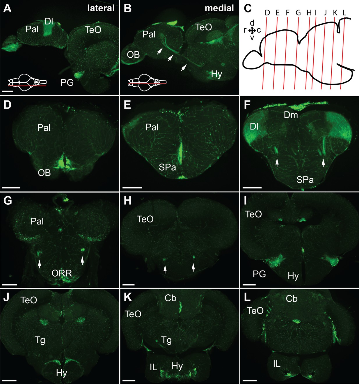
GFP expression of the Tg(279A-GFP) adult brain.
(A and B) Sagittal sections of the Tg(279A-GFP) adult brain (anterior to the left). A is a more lateral section showing the PG and Dl, and B is a more medial section showing the PG axons coursing to the Dl (arrows). (C) Schematic drawing of a sagittal section indicating the level of the frontal sections shown in (D–L). (D–L) Frontal sections of the Tg(279A-GFP) adult brain from anterior to posterior. The PG-pallial projection fibers are indicated with arrows in (F), (G), and (H). Note that the GFP expression is not exclusive to the PG-pallial projection neurons, but some other structures such as the olfactory bulb (OB) and the hypothalamus (Hy) are also labeled. Abbreviations: Cb, cerebellum; Dl, lateral part of dorsal telencephalic area; Dm, medial part of dorsal telencephalic area; IL, inferior lobe; Hy, hypothalamus; OB, olfactory bulb; ORR, optic recess region; Pal, pallium; PG, preglomerular complex; SPa, subpallium; TeO, optic tectum; Tg, tegmentum. Brain orientation: r, rostral; c, caudal; d, dorsal; v, ventral. Scale bars,=200 µm.