
Fig. 6
- ID
- ZDB-IMAGE-200928-10
- Publication
- Bloch et al., 2020 - Non-thalamic origin of zebrafish sensory nuclei implies convergent evolution of visual pathways in amniotes and teleosts
- All Figures
- Figures for Bloch et al., 2020
|
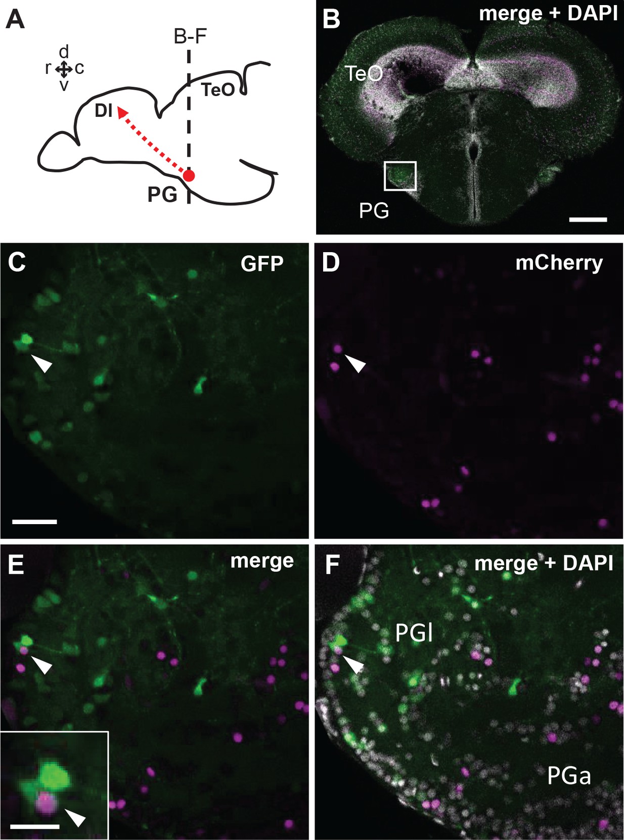
Fig. 6
Co-localization of GFP and mCherry in the adult PG cells of the quadruple transgenic line Tg(her5:ERT2CreERT2;βactin:lox-stop-lox-hmgb1-mCherry;279A-GFP) following the tamoxifen induction at 24 hpf.
(A) Schematic drawing of the adult zebrafish brain indicating the level of section through the PG shown in B-F. (B) A single plane confocal image showing a global view of the frontal section of a 3 mpf zebrafish brain. The white square indicates the PGl shown in C-F at a higher magnification. (C–F) A confocal image (5 µm projection) showing the co-localization of GFP and mCherry in PGl (arrowheads). Inset of E shows the double-labeled cell at a higher magnification. Abbreviations: Dl, lateral part of dorsal telencephalic area; PG, preglomerular complex; PGa, anterior preglomerular nucleus; PGl, lateral preglomerular nucleus; TeO, optic tectum. Brain orientation: r, rostral; c, caudal; d, dorsal; v, ventral. Scale bars = 200 µm (B); 30 µm (C; applicable to D-F); 10 µm (inset of E).