
IMAGE
Fig. 4
- ID
- ZDB-IMAGE-200924-1
- Publication
- Zhu et al., 2019 - Zebrafish prmt5 arginine methyltransferase is essential for germ cell development
- All Figures
- Figures for Zhu et al., 2019
Image
|
Figure Caption
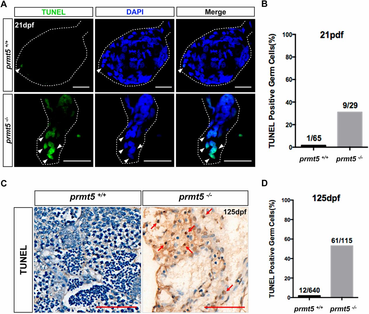
Fig. 4 Deletion of prmt5 in zebrafish leads to germ cell apoptosis. (A) Apoptotic cells in the gonads (white dashed lines) of prmt5+/+ and prmt5−/− zebrafish at 21 dpf. More apoptotic cells (green; white arrowheads) are found in the prmt5−/− zebrafish gonads than in prmt5+/+ zebrafish gonads (n=4, respectively). (B) The ratios of apoptotic cells in prmt5+/+ and prmt5−/− zebrafish at 21 dpf. (C) Apoptotic cells (arrows) in the gonads of prmt5+/+ and prmt5b−/− zebrafish at 125 pdf; n=4, respectively. (D) The ratios of apoptotic cells in prmt5+/+ and prmt5−/− zebrafish at 125 dpf. Scale bars: 20 μm.
Figure Data
Acknowledgments
This image is the copyrighted work of the attributed author or publisher, and
ZFIN has permission only to display this image to its users.
Additional permissions should be obtained from the applicable author or publisher of the image.
Full text @ Development