
Fig. S2
- ID
- ZDB-IMAGE-200910-30
- Publication
- Martorano et al., 2019 - The zebrafish orthologue of the human hepatocerebral disease gene MPV17 plays pleiotropic roles in mitochondria
- All Figures
- Figures for Martorano et al., 2019
|
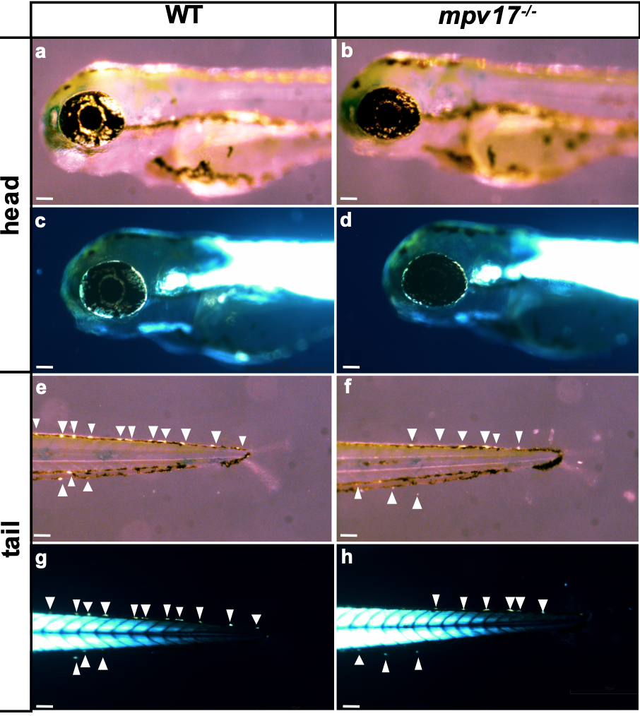
Fig. S2
Comparison between different counting methods of iridophores in wild-type and mpv17-/- larvae at 3 dpf. Bright-field pictures (a, b, e, f), acquired according to Krauss and colleagues’ method: the angle of illumination had been adjusted individually to allow optimal visualization of iridophores (Krauss et al., 2013). Birefringence images (c, d, g, h), acquired by using a polarizer and an analyser lens, according to previously published methods (Smith et al., 2013). Representative images of iridophores along the tail (g, h) or off the eye (c, d) were taken without the need of modifying the incident light and by completely abolishing background. The arrows point to iridophores. Bars, 100 μm.