
Fig. 5
- ID
- ZDB-IMAGE-200902-14
- Genes
- Publication
- Peskin et al., 2020 - Notochordal Signals Establish Phylogenetic Identity of the Teleost Spine
- All Figures
- Figures for Peskin et al., 2020
|
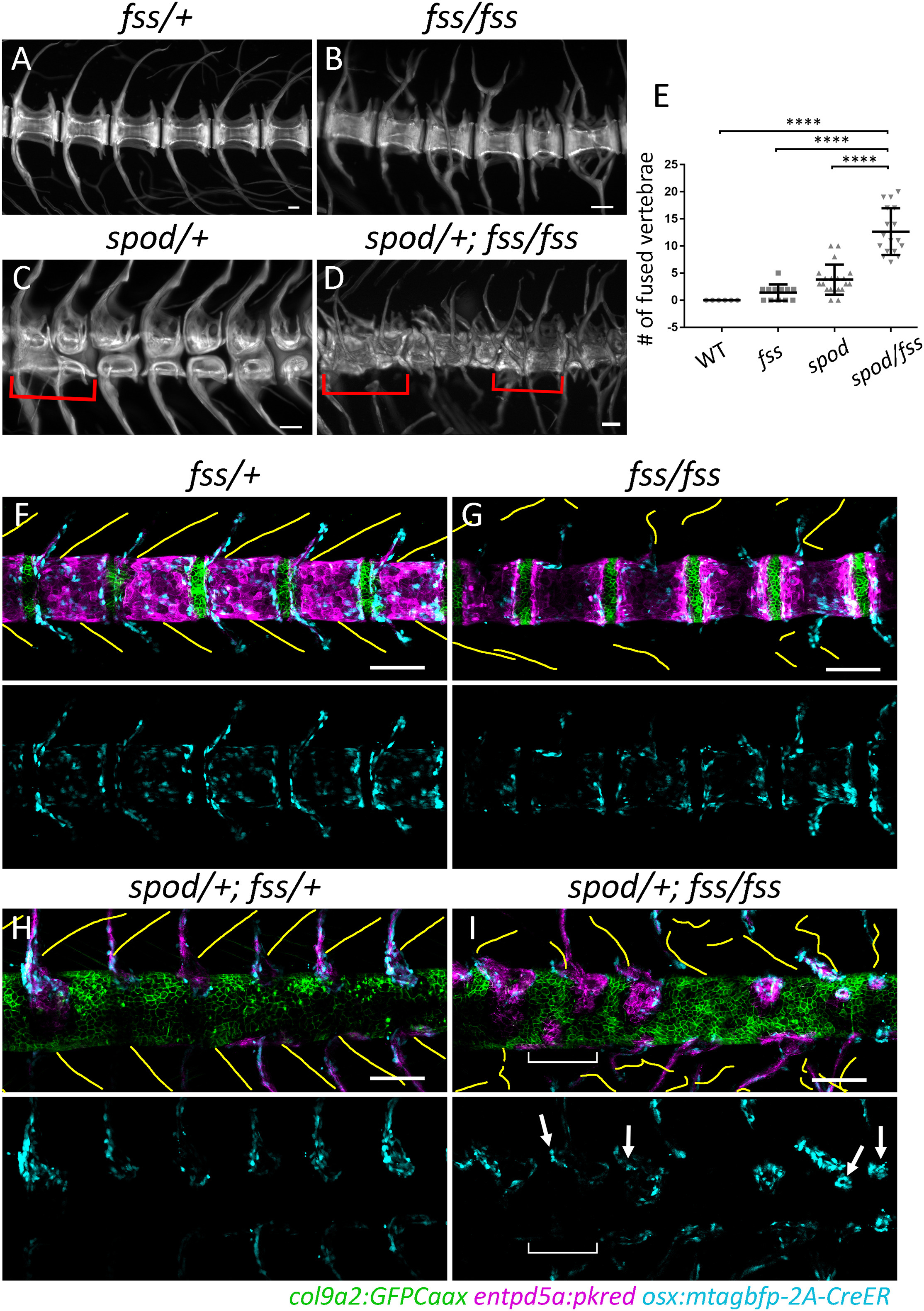
Fig. 5 Sensitization of cmnspod Mutants to Tetrapod Segmentation Programs (A–D) Alizarin red stained skeletal preparations at 6 wpf. (A) Wild-type skeletal preps demonstrate meristically patterned autocentra and arches. (B) tbx6fss/fss mutants display irregular arch morphology and patterning, while centra remain regularly segmented. (C) cmnspod/+ mutants have the opposite phenotype wherein arch patterning is normal and the centra contain large vertebral clefts. (D) Double mutants in which both notochord and somite segmentation have been disrupted are not simply a combination of the two aforementioned phenotypes. In contrast, double mutants form highly irregular vertebral structures with very little discernable patterning. Red brackets indicate vertebral fusion events. (E) The number of fused vertebrae in each condition was quantified along the body axis using alizarin red stained skeletal preparations. The number of fused vertebrae in double mutants was significantly greater than in all other conditions. Wild-type n = 6; tbx6fss/fss n = 12; cmnspod/+ n = 20; cmnspod/+;tbx6fss/fss n = 17 fish. Error bars indicate standard deviation. **** indicates p value less than 0.0001. (F–I) Alteration In notochordal patterning demonstrating alteration in early sensitivity of spondo mutants to tbx6 mutations; yellow lines indicate traced somite segment boundaries; blue channel (osteoblasts) has been isolated below each composite image. (F and H) In wild-type and spondo fish, regions of somite boundaries correlate with articulated pattern of vertebral arches. (G) The tbx6fss/fss mutant has impaired somite segmentation where partial segment boundaries form without any metameric pattern. Lack of paraxial mesoderm patterning alters the morphology and arrangement of developing vertebral arches while notochord segmentation is largely unaffected. There is some variability in size of the centra domains (not quantified). (I) In spondo;tbx6 double mutants, both notochord and somite segmentation are impaired. Osteoblasts are found nearby partial boundaries. The bracket indicates a region devoid of segment boundaries as well as osteoblasts. Arrows mark osteoblasts in close proximity to partial somite boundaries that have begun to spill over to form hemicentra structures.