
Image
|
Figure Caption
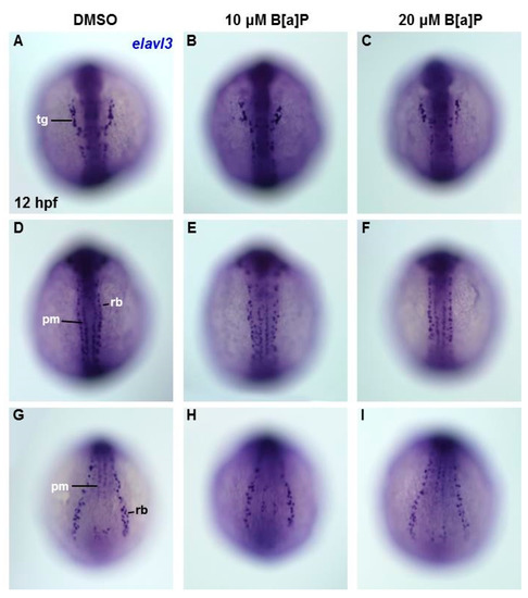
Fig. 4
Lack of obvious neural cell defects in B[a]P-treated embryos at 12 hpf. In situ hybridization staining with a probe against elavl3 was performed to analyze the early development of neural cells (A–I); all images show the dorsal view. Images in (A–C) are focused on the anterior trunk; (D–F) are focused on mid-trunk region and (G–I) are focused on the tail region. tg, trigeminal ganglion; pm, primary motor neurons; rb, Rohon–Beard cells.
Acknowledgments
This image is the copyrighted work of the attributed author or publisher, and
ZFIN has permission only to display this image to its users.
Additional permissions should be obtained from the applicable author or publisher of the image.
Full text @ Antioxidants (Basel)