
Fig. 5
- ID
- ZDB-IMAGE-200820-17
- Publication
- El-Nachef et al., 2020 - De novo enteric neurogenesis in post-embryonic zebrafish from Schwann cell precursors rather than resident cell types
- All Figures
- Figures for El-Nachef et al., 2020
|
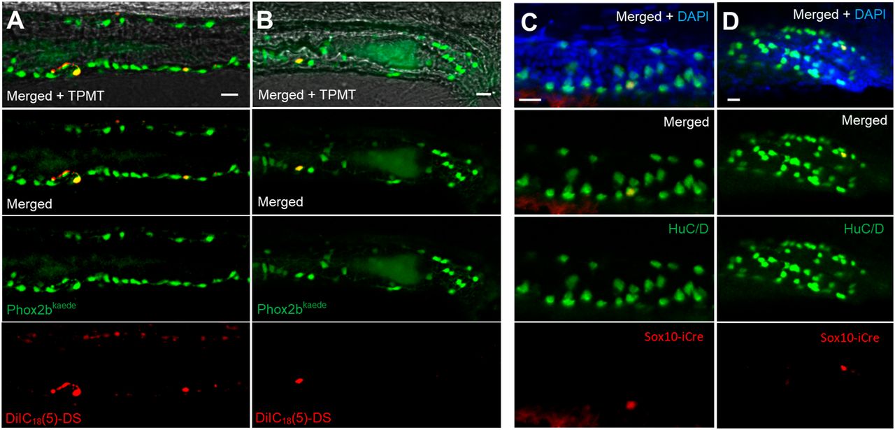
Fig. 5 Lineage tracing demonstrates a trunk neural crest origin of post-embryonic neurons. (A,B) Phox2b-kaede embryos underwent neural tube injections of a far-red lipophilic dye at 30 hpf, after the vagal crest has delaminated from the neural tube. Live images at 6 dpf of the midgut (A) and hindgut (B) demonstrate Phox2b-kaede enteric neurons that colocalize with the dye, indicating their trunk origin. (C,D) Fish from the inducible Sox10-Cre line were exposed to 4-OHT at 3.5 dpf and underwent IHC for the Cre reporter, mCherry and the neuronal marker HuC/D at 5.5 dpf, with Cre-labelled enteric neurons observed in the midgut (C) and hindgut (D). As Cre induction occurring after Sox10 is no longer present within the intestine, these results support a trunk neural crest origin of these enteric neurons. Scale bars: 20 µm.