
Fig. 4
- ID
- ZDB-IMAGE-200820-16
- Publication
- El-Nachef et al., 2020 - De novo enteric neurogenesis in post-embryonic zebrafish from Schwann cell precursors rather than resident cell types
- All Figures
- Figures for El-Nachef et al., 2020
|
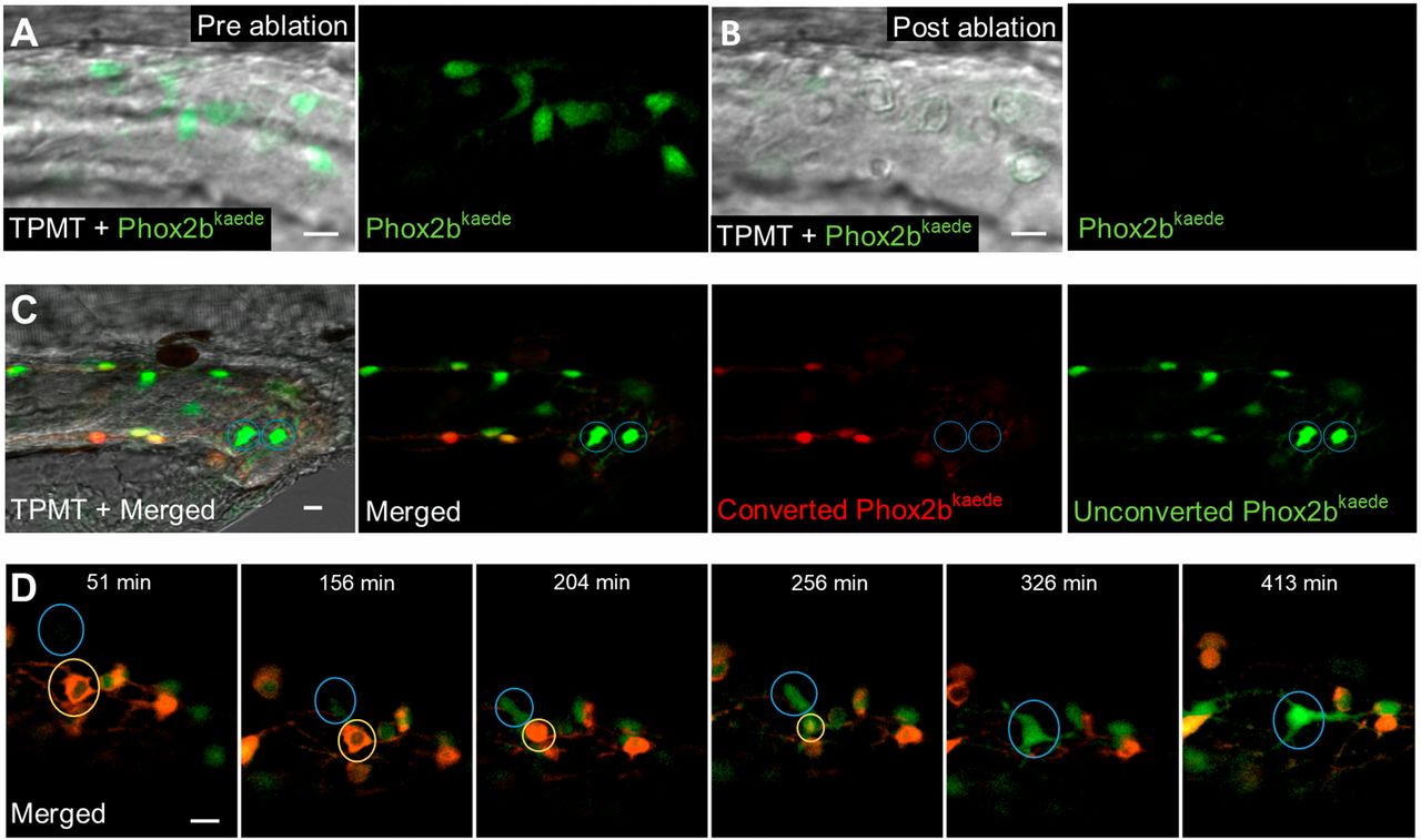
Fig. 4 De novo enteric neurons replace ablated neurons in a post-embryonic injury model. (A,B) Prior to two-photon laser ablation, Phox2b-kaede enteric neurons are clearly visualized within the hindgut (A). After ablation, these neurons are no longer present in the hindgut, and TPMT reveals the injury site to be restricted to the neuron location (B). (C) At 4.5 dpf, fish underwent laser ablation of ten enteric neurons within the distal hindgut, followed by photoconversion of all remaining enteric neurons within the whole length of the gut. Live imaging was performed 12 h later and detected multiple de novo green fluorescent-only enteric neurons in the hindgut. (D) 8 h time-lapse of a fish at 4.5 dpf that underwent focal injury (but not complete ablation) of enteric neurons followed by pan-gut photoconversion reveals the involution of an injured neuron (yellow circle) that is replaced by a de novo green fluorescent-only enteric neuron. The new neuron (blue circle) initially appears very faintly at the dorsalmost aspect of the intestine but increases in intensity as it migrates to replace the involuted neuron and extends projections to neighbouring neurons. Scale bars: 10 µm.