
Fig. 2
- ID
- ZDB-IMAGE-200813-2
- Publication
- Lex et al., 2020 - Telomere shortening produces an inflammatory environment that increases tumor incidence in zebrafish
- All Figures
- Figures for Lex et al., 2020
|
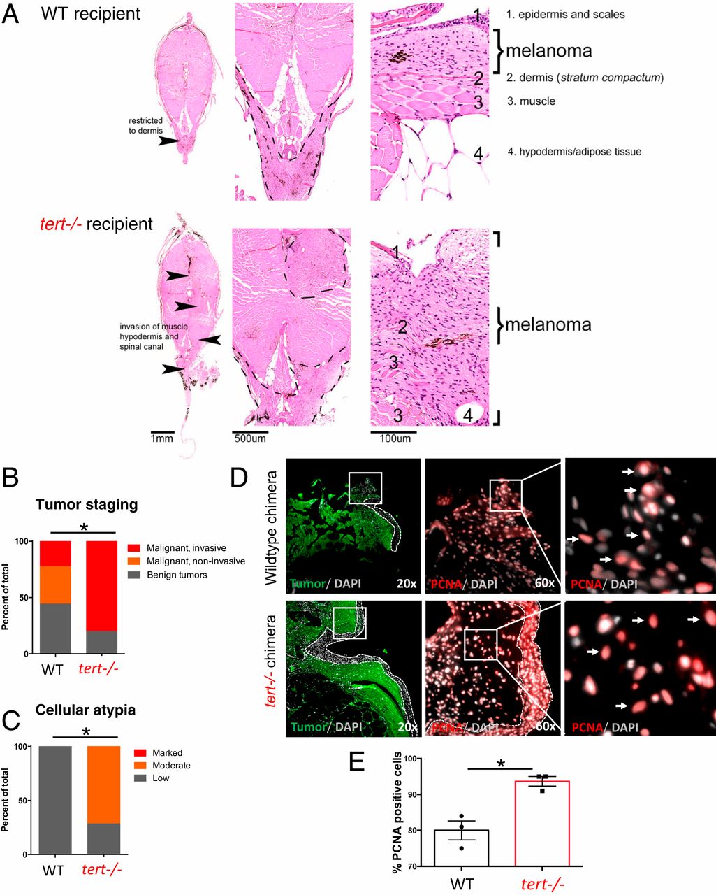
Fig. 2 tert−/− tissues increase melanoma invasiveness and progression. (A) Hematoxylin and eosin (H and E) images of melanoma arising in a WT (Upper) or tert−/− recipient fish. Strong infiltration into other tissues was typical in tert−/− fish but not in WT (arrowheads). (B) Melanomas were staged according to histopathology into benign lesions (melanosis), noninvasive and invasive malignant tumors (P = 0.0269; WT n = 9; tert−/− n = 10). (C) Analysis of malignant tumors for cellular atypia (P = 0.0278; WT n = 5; tert−/− n = 7). (D) Representative immunofluorescence images of PCNA-positive cells (red) in melanoma (green) developed in WT chimeras and in tert−/− chimeras. Dashed lines locate the skin (no green fluorescence), squares show the place of amplification, and arrows indicate PCNA-positive cells. (E) Quantification of PCNA-positive melanoma cells in WT and tert−/− chimeras. Data are represented as mean ± SEM. *P < 0.05; n = 3.