
Fig. 5
- ID
- ZDB-IMAGE-200812-17
- Publication
- Davis et al., 2020 - Chloroquine kills hair cells in zebrafish lateral line and murine cochlear cultures: Implications for ototoxicity
- All Figures
- Figures for Davis et al., 2020
|
Fig. 5
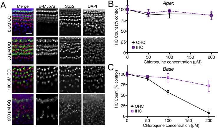
Chloroquine exposure causes dose-dependent hair cell loss in neonatal mouse organ of Corti cultures. (A) Fluorescent imaging of basal turn cultures treated with 0, 50, 100, and 200 μM chloroquine and labeled with antibodies specific to hair cells (Myosin Vlla), or organ of Corti supporting cells (Sox2), or all nuclei (DAPI). Note that 50 μM chloroquine exposure (row labeled 50 μM CQ) did not reveal clear loss or disruption of any cell type (compare with top row (0 μM CQ). However, 100 μM and 200 μM chloroquine exposure resulted in moderate and extreme loss of outer hair cells, respectively. Conversely, there was no obvious loss of other cell types at any treatment condition. (B) Apical coil cultures demonstrate little or no loss of inner or outer hair cells following treatment with chloroquine at any of the concentrations used (p = 0.6087). (C) Basal coil hair cells, however, showed significantly decreased viability when treated with chloroquine (p < 0.0001), and outer hair cells in the base were more dramatically affected by drug exposure than inner hair cells (F = 26.11, p < 0.0001). Error bars represent ±1 standard deviation. Scale bar = 20 μm.
Reprinted from Hearing Research, 395, Davis, S.N., Wu, P., Camci, E.D., Simon, J.A., Rubel, E.W., Raible, D.W., Chloroquine kills hair cells in zebrafish lateral line and murine cochlear cultures: Implications for ototoxicity, 108019, Copyright (2020) with permission from Elsevier. Full text @ Hear. Res.