
Image
|
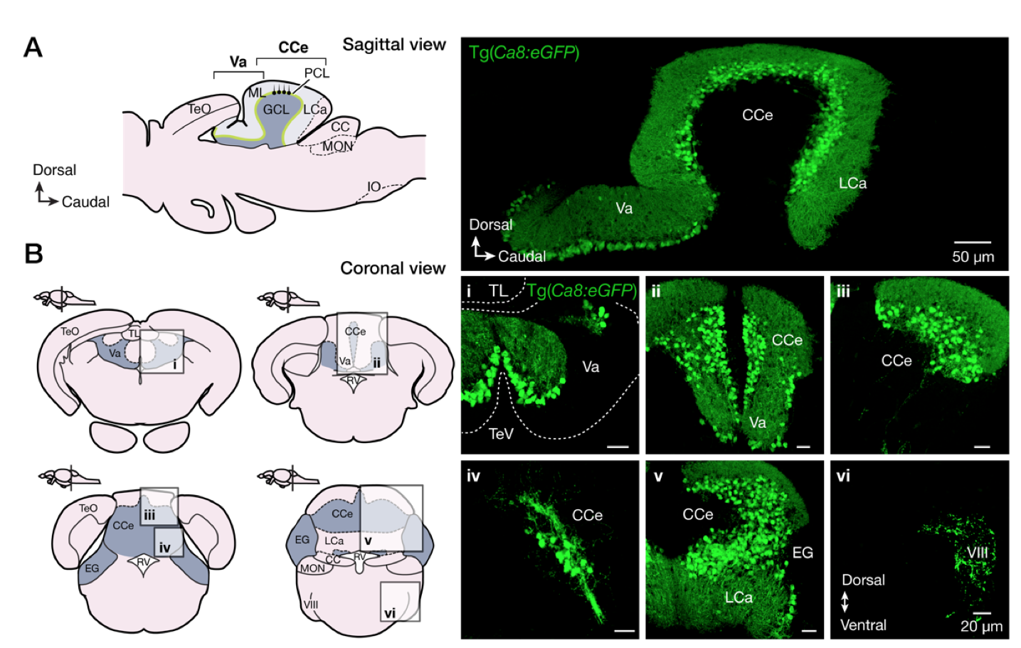
Figure Caption
Fig. S2
Expression pattern of Ca8:eGFP in the adult zebrafish brain. (A-B) Sagittal and coronal sections are showing the specificity of the eGFP expression in all Purkinje cells (green) of the adult zebrafish cerebellum. CC, crista cerebellaris, CCe, corpus cerebelli; EG, eminentia granularis; GCL, granule cell layer; IO, oliva inferior; LCa, lobus caudalis cerebelli; ML, molecular layer; MON, medial octavolateralis nucleus; PCL, Purkinje cell layer; RV, rhombencephalic ventricle; TeO, tectum opticum; Tev, tectal ventricle; Va, valvular cerebelli; VIII, octaval nerve.
Acknowledgments
This image is the copyrighted work of the attributed author or publisher, and
ZFIN has permission only to display this image to its users.
Additional permissions should be obtained from the applicable author or publisher of the image.
Full text @ Proc. Natl. Acad. Sci. USA