
Fig. 8
- ID
- ZDB-IMAGE-200728-47
- Publication
- Brandt et al., 2020 - Core Hippo pathway components act as a brake on Yap/Taz in the development and maintenance of the biliary network
- All Figures
- Figures for Brandt et al., 2020
|
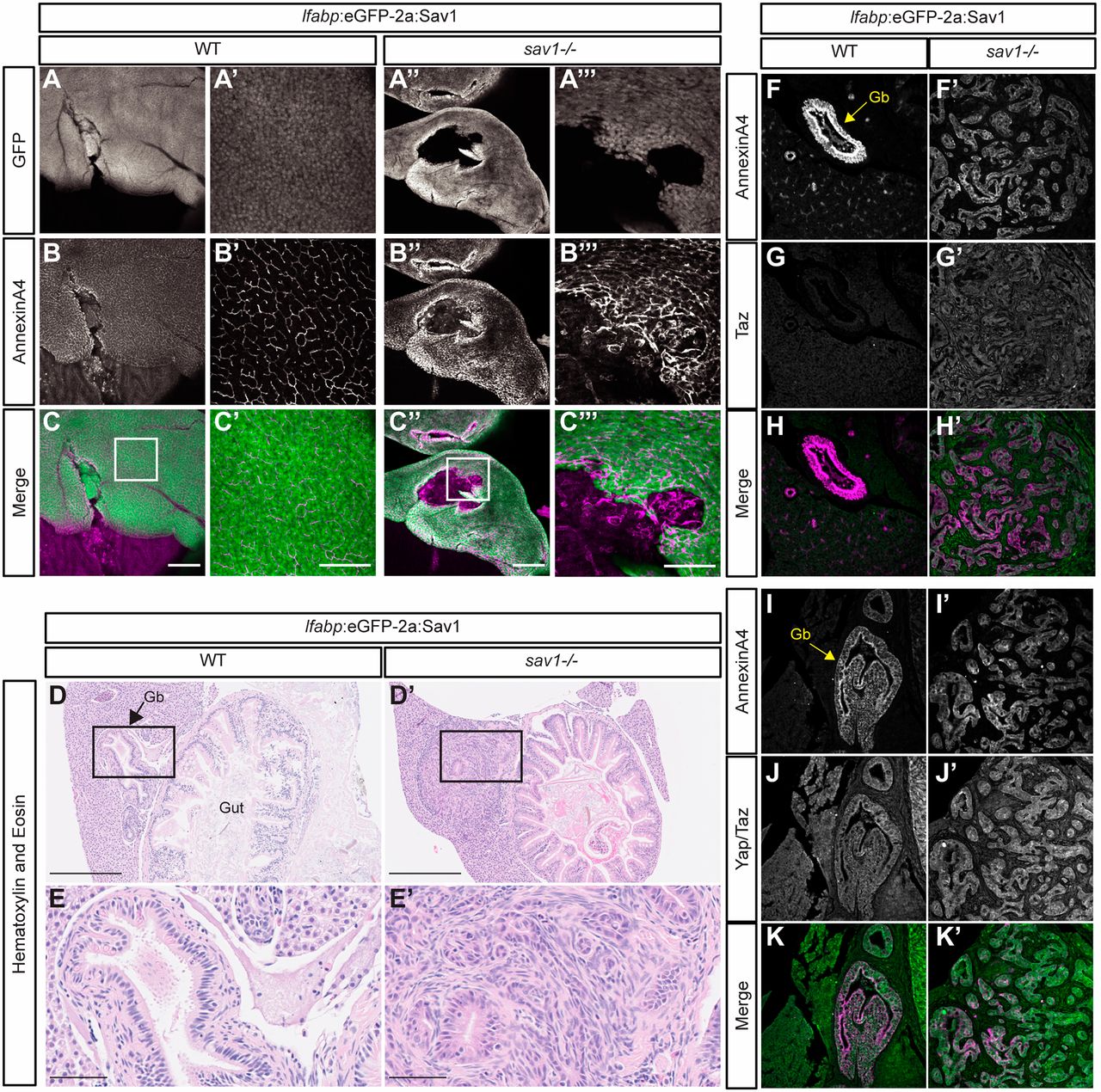
Fig. 8 Hippo signaling functions cell-autonomously in the maintenance of the gallbladder and extrahepatic biliary ducts. (A-A‴) Whole-mount GFP staining in wild-type and sav1−/− adult fish. (B-B‴) Whole-mount annexin A4 staining in wild-type and sav1−/− adult fish. (C-C‴) Merged images of GFP (green) and annexin A4 (magenta). Scale bars: 250 μm in C,C″; 100 μm in C′,C‴. (D-E′) Hematoxylin and Eosin histology of the same fish analyzed in A-C‴. Gb, gallbladder (n=10 wild type, lfabp:eGFP-2a:Sav1+; n=8 sav1−/−, lfabp:eGFP-2a:Sav1+). Scale bars: 250 µm in D and E; 50 µm in D′ and E′. (F,F′,I,I′) Annexin A4 immunostaining on wild-type and sav1−/− dissected liver and gut of fish carrying lfabp:eGFP-2a:Sav1 transgene. Gb, gallbladder. (G,G′) Taz staining on the same sections shown in F,F′. (J,J′) Yap and Taz staining on the same sections shown in I,I′. (H,H′,K,K′) Merged images of Taz alone, Yap and Taz (green), and annexin A4 (magenta) (n=5 wild type, lfabp:eGFP-2a:Sav1+, n=3 sav1−/−, lfabp:eGFP-2a:Sav1+).