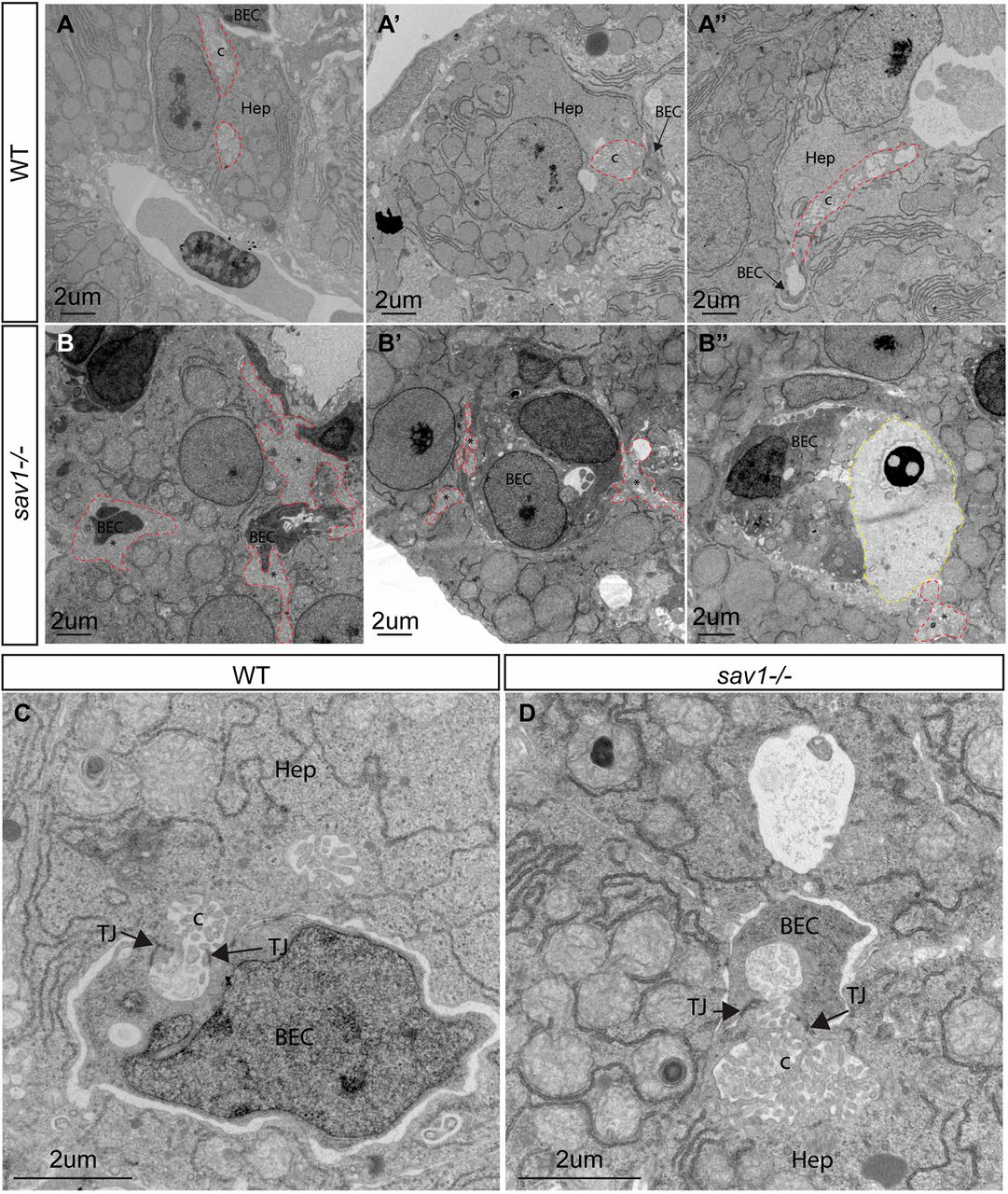
Fig. 6 Ultrastructural defects of bile duct canaliculi in sav1−/− larvae. (A-A″) TEM examples of wild-type canaliculi and hepatocyte-biliary epithelial cell junctions. Canaliculi are outlined with dashed red lines. (B-B″) sav1−/− siblings of A-A″ showing examples of hepatocyte-to-biliary cell borders that contain membranous material reminiscent of canaliculi, but lack normal structure (dashed red lines). In B″, a dashed yellow line outlines excessive extracellular space filled with electron-dense material. (C) Higher magnification of wild-type hepatocyte-biliary epithelial cell junction and canaliculi. (D) Example of a rare canaliculi present in sav1−/− larvae showing shorter length and increased diameter. Hep, hepatocyte; BEC, biliary epithelial cell; c, canaliculi; TJ, tight junction. Asterisks indicate abnormal canaliculi or abnormal hepatocyte membrane. n=3 wild type, n=3 sav1−/−. All images are of 10 dpf larvae.
Image
Figure Caption
Figure Data
Acknowledgments
This image is the copyrighted work of the attributed author or publisher, and
ZFIN has permission only to display this image to its users.
Additional permissions should be obtained from the applicable author or publisher of the image.
Full text @ Development

