
|
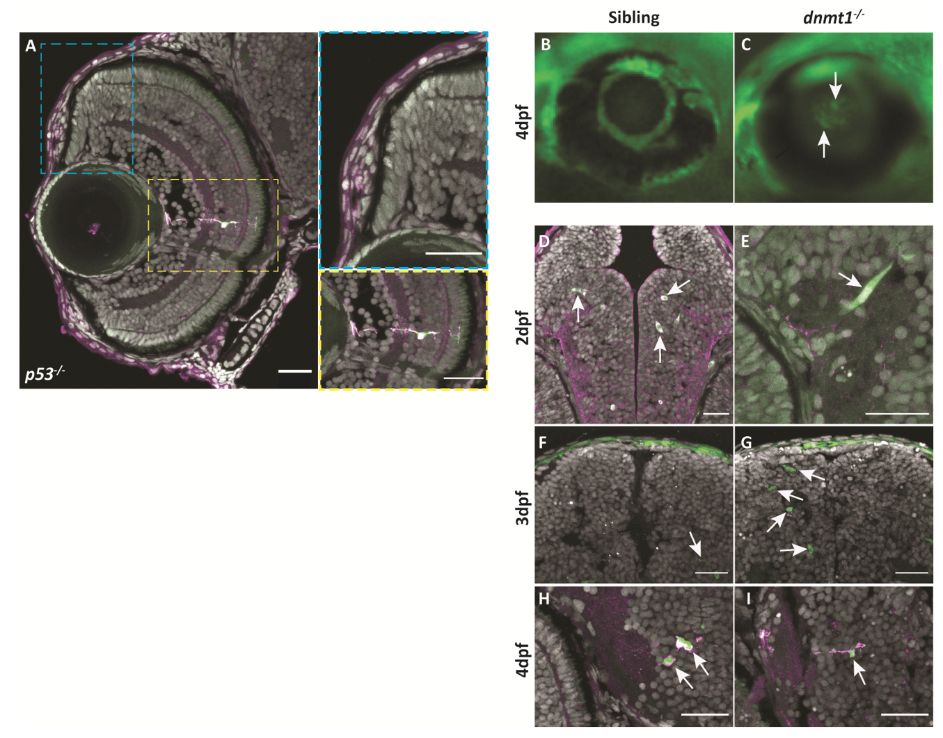
Figure S5
L1RE3-EGFP transgene expression is more prominent in dnmt1-/- larvae. A-A’’. Transverse section of Tg(CMV:Hsa.L1RE3,EGFP,myl7:EGFP;p53-/-) larvae at 4dpf. A. L1RE3-EGFP+ retinal cells labeled with endogenous EGFP. Cyan (A‘) and yellow (A’‘) dotted boxes indicate magnified images to the right of panel A. A’. Magnified image of Tg(CMV:Hsa.L1RE3,EGFP,myl7:EGFP;p53-/-) CMZ showing no expression of the L1RE3-EGFP transgene. A’’. Magnified image of retinal neuron expressing L1RE3-EGFP transgene. B-I. All images are taken from Tg(CMV:Hsa.L1RE3,EGFP,myl7:EGFP) larvae that are either dnmt1+/+ (B,D,F,H) or dnmt1-/- (C,E,G,I). B-C. Whole-mount images of 4dpf eyes demonstrating L1RE3-EGFP transgene activation seen through the lens of dnmt1-/- larvae and not siblings. D,F,H. Sibling larvae expressing the L1RE3-EGFP transgene in the brain. E,G,I. dnmt1-/- larvae expressing the L1RE3-EGFP transgene in the brain. Nuclei labeled with DAPI (gray). Endogenous L1RE3-activated EGFP labeled in green. EGFP antibody is magenta. Scale bars: 30 μm in all images. Dorsal is up in all images.