
Figure 1
- ID
- ZDB-IMAGE-200725-21
- Publication
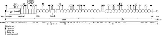
- Toms et al., 2020 - Clinical and preclinical therapeutic outcome metrics for USH2A-related disease
- All Figures
- Figures for Toms et al., 2020
|
Figure 1
Position and type of