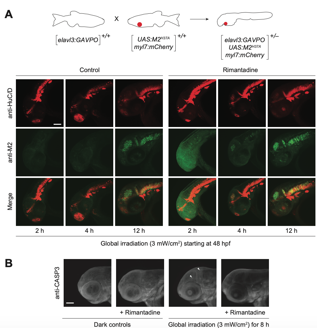
Fig. S5
Light-inducible, neuron-specific M2H37A expression using the GAVPO system. (A) Tg(elavl3:GAVPO; UAS:M2H37A;myl7:mCherry) embryos were globally irradiated with blue LED light at 48 hpf for the designated durations and cultured in the presence or absence of rimantadine. The embryos were then fixed, and immunostained for M2 and HuC/D expression. Representative confocal micrographs are shown as maximum intensity projections of 39 to 51 z-stack images (5-μm optical sections), demonstrating overlaying domains of M2H37A and HuC/D expression in the anterior CNS. (B) Tg(elavl3:GAVPO;UAS:M2H37A;myl7:mCherry) embryos were globally irradiated with blue LED light at 48 hpf for 8 h and cultured in the presence or absence of rimantadine. The embryos were fixed 12 h later, and immunostained for activated caspase-3. Representative confocal micrographs are shown as maximum intensity projections of 30 to 50 z-stack images (7-μm optical sections), demonstrating limited non-cell autonomous apoptosis. Arrowheads: cells with activated caspase-3. Embryo orientations: lateral view, anterior left. Scale bars: 1-0 μm.

