
Figure 3
- ID
- ZDB-IMAGE-200720-17
- Genes
- Publication
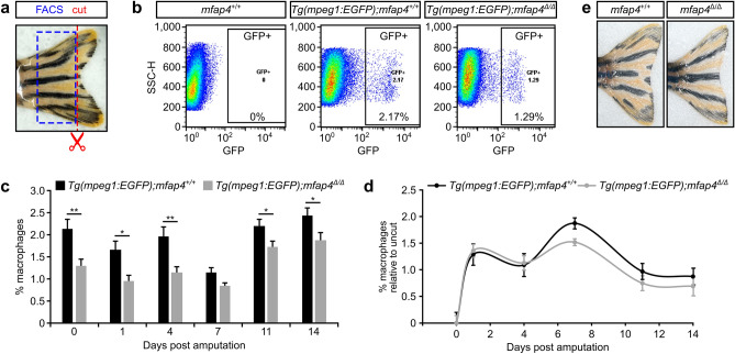
- Ong et al., 2020 - Microfibril-associated glycoprotein 4 (Mfap4) regulates haematopoiesis in zebrafish
- All Figures
- Figures for Ong et al., 2020
|
Figure 3