YPEL3 is an evolutionarily conserved disease-causing candidate gene.
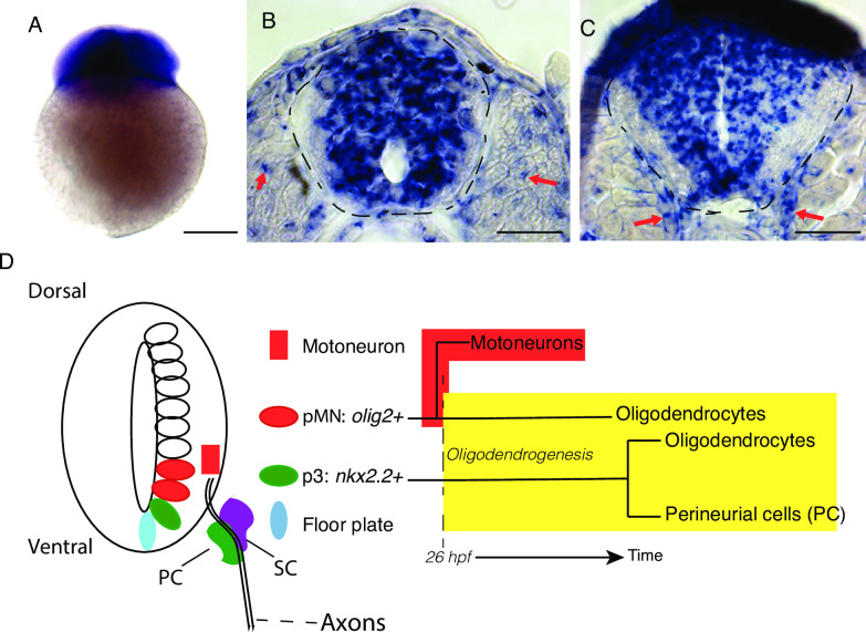
(A-C) ypel3 is maternally deposited and expressed during neural development in zebrafish. (A) Two-cell stage embryo. (B-C) Transverse sections. (B) At 24 hours postfertilization (hpf), ypel3 is expressed broadly in the spinal cord and sparsely in the somites (red arrows). (C) At 56 hpf, ypel3 is expressed in the spinal cord and in the region where the motor roots develop (red arrows). (D) Diagram of spinal cord domains, their derivatives, and molecular markers. The pMN domain (olig 2+) gives rise first to motoneurons, and then later oligodendrocytes (Briscoe et al., 2000; Jessell, 2000; Danesin and Soula, 2017). The p3 domain (nkx2.2+) is localized ventral to the pMN domain and gives rise to oligodendrocytes and a subset of perineurial cells (Clark et al., 2014; Kucenas et al., 2008b). Schwann cells (SC) are neural crest derivatives. Scale bar: 250 μm in A, 25 μm in B and C.
This image is the copyrighted work of the attributed author or publisher, and
ZFIN has permission only to display this image to its users.
Additional permissions should be obtained from the applicable author or publisher of the image.
Full text @ PLoS Genet.
Your Input Welcome
Thank you for submitting comments. Your input has been emailed to ZFIN curators who may contact you if
additional information is required.
Oops. Something went wrong. Please try again later.