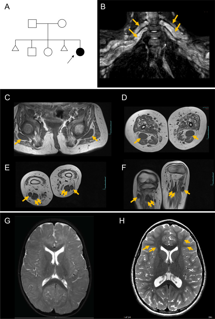
Proband has myelination defects and greatly enlarged nerves.
(A) Pedigree of the family; arrow points to the proband; triangular shapes indicate instances of spontaneous abortion. (B-F) MRI of upper and lower extremities demonstrates enlarged nerves. (B) Image showing greatly enlarged brachial plexus. Arrows indicate the thick peripheral nerves. (C) Sciatic nerve at the level of the femoral head (arrows). (D) Sciatic nerve at the level of the mid-thigh (arrows). (E) Posterior tibial (double arrows) and peroneal (single arrows) nerves at their origins. (F) Posterior tibial (double arrows) and peroneal (single arrows) nerves at the level of the fibular head. Scale bars, 3 cm. (G) Representative brain MRI (axial, T2-weighted) showing poor delineation of white and gray matter, indicating reduced myelination, when compared to age-matched control (H). Note the delineation of white and gray matter in control (arrows) that is not seen in the patient.
Acknowledgments
This image is the copyrighted work of the attributed author or publisher, and
ZFIN has permission only to display this image to its users.
Additional permissions should be obtained from the applicable author or publisher of the image.
Full text @ PLoS Genet.
Your Input Welcome
Thank you for submitting comments. Your input has been emailed to ZFIN curators who may contact you if
additional information is required.
Oops. Something went wrong. Please try again later.