
|
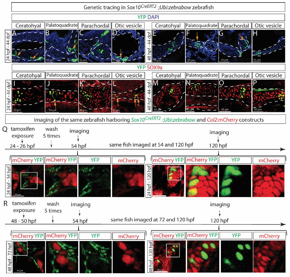
Fig. s9 SCPs and Schwann cells give rise to craniofacial chondrocytes during the postnatal development of zebrafish. (A-P) Sox10CreERT2;Ubi:zebrabow zebrafish were exposed to 4OH-tamoxifen for 2 hours either at 24 hpf (A-D and I-L) or 48 hpf (E-H and M-P) and analyzed at 44 days post fertilization (dpf). Progeny of genetically-labeled SCPs were detected in various cartilage elements including ceratohyal (A, E, I, M), palatoquadrate (B, F, J, N), parachordal (C, G, K, O) and otic vesicle (D, H, L, P). Cartilage elements were visualized either by characteristic morphological appearance of DAPI-counterstained sections (A-H) or SOX9a (I-P). Orange arrows in A, B, D, F, G, J, L indicate YFP+ perichondrial cells. (Q) The Sox10CreERT2;Ubi:zebrabow zebrafish strain was crossed with the Col2:mCherry strain to visualize chondrogenesis simultaneously with genetic tracing of SCPs, exposed to 4OH-tamoxifen at 24-26 hpf and imaged live at 54 hpf followed by the same fish imaging at 120 hpf. No overlap between SCPs and cartilage cells was observed initially, but at 120 hpf. (R) The same experiment as in Q but with different initial labeling and detection time-points as indicated. Images in Q-R are single confocal slices of the stained samples.