
Image
|
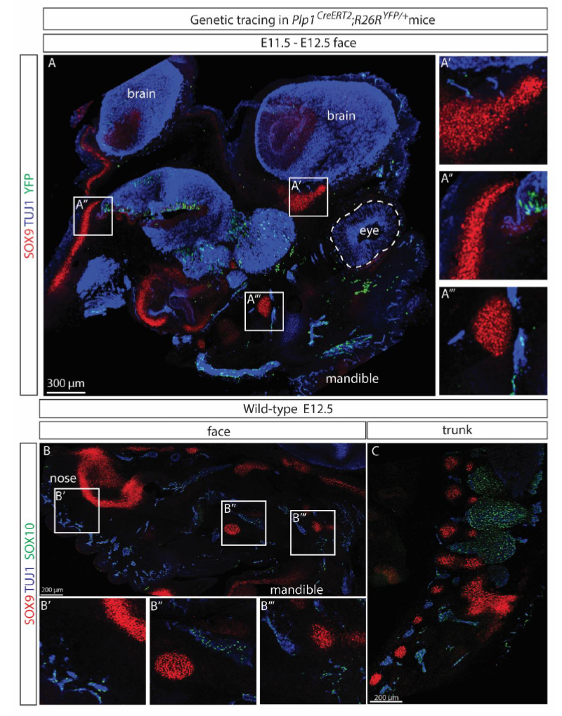
Figure Caption
Fig. s3 (A) A representative confocal scan of the entire face of an Plp1CreERT2;R26RYFP/+ embryo traced from E11.5 to E12.5 showed no overlap between YFP+ SCPs and chondro-progenitors revealed by immunostaining for SOX9. Nerves were visualized by TUJ1 immunostaining. (B-C) SOX9+ and SOX10+ cells were distinct populations on embryonic day E12.5 in both the facial region (B) and trunk (C, spine area shown) of wild-type C57BL/6J mice.
Acknowledgments
This image is the copyrighted work of the attributed author or publisher, and
ZFIN has permission only to display this image to its users.
Additional permissions should be obtained from the applicable author or publisher of the image.
Full text @ Proc. Natl. Acad. Sci. USA