
Fig. 8
- ID
- ZDB-IMAGE-200618-20
- Publication
- Hehr et al., 2018 - Polarity and morphogenesis of the eye epithelium requires the adhesion junction associated adaptor protein Traf4
- All Figures
- Figures for Hehr et al., 2018
|
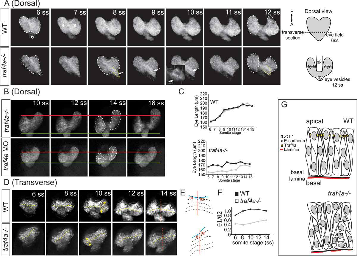
Fig. 8 Eye vesicle evagination and elongation abnormal with Traf4a loss. Sequential confocal maximal projection of optical sections from Tg(rx3:GFP) control, e1i1 MO injected and traf4a-/- eye vesicles over the period of eye evagination and elongation (6–18 ss). Time-lapse sequences were obtained for the dorsal view from n = 6 control, n = 3 morphant and n = 5 traf4a-/- embryos, and for the transverse view from n = 8 control, n = 9 morphant and n = 3 traf4a-/- embryos. A-B: Dorsal view, from the 6–12 ss (A) and from the 10–16 ss (B). Eye vesicles outlined in white, and a bulge in the eye field (ef) outlined in yellow, and shown as a 3-D reconstruction (inset). Asymmetric evagination of the eye vesicles indicated by arrows. In B, the green and red lines mark the anterior and posterior extent of the left eye vesicles at the 10 ss. C: Length of the two eye vesicles over time for the two embryos shown in A. D-F: Transverse view (D), from the 6–14 ss. Dotted yellow lines mark the ventricles, and the red line the midline of the neural keel. These lines are represented anew in E, and the ratios of the angles of the ventricle to the midline for the two eye vesicles over time are shown in F. G: Model for Traf4a function in the eye epithelium. Traf4a is an adaptor protein found at the plasma membrane that associates with both adherens (E-cadherin) and tight (ZO-1) junction proteins in other systems. Based on the literature, we propose that Traf4a is co-localized with these proteins at the apical surface of the nascent zebrafish eye epithelium. In the absence of Traf4a, ZO-1 is present in a patchy fashion on the apical surface and ectopic to the apical surface, while E-Cadherin does not accumulate apically. Further, expression of the basally located protein Laminin is disrupted. Consequently a disorganized epithelium is formed, whose cells can lose their apical attachment and radially-oriented nuclei, which is associated with a failure of eye vesicles to evaginate and elongate