IMAGE

Fig. 6

ID
ZDB-IMAGE-200615-8
Source
Figures for Wang et al., 2020
Image
Figure Caption

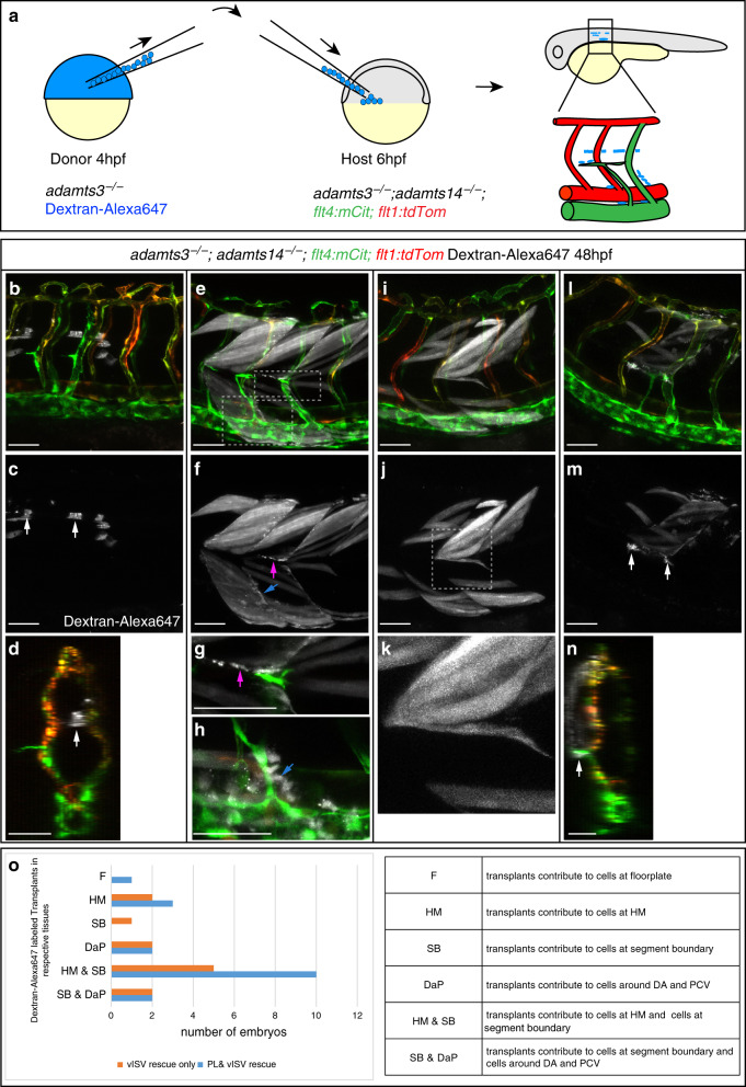
Fig. 6 <italic>adamts14</italic>-expressing floorplate and mesenchymal cells can rescue venous sprouting and PL formation in <italic>adamts3</italic>; <italic>adamts14</italic> double mutants.

a Scheme outlining the transplantation assay: embryos homozygous mutant for adamts3 were injected with Dextran-Alexa647 to mark the donor cells. At 4 hpf, cells from the donor were transplanted into adamts3; adamts14 double mutant host embryos (6 hpf). Host embryos were subsequently analyzed for rescue of venous sprouting and the location of the transplanted material at 48 hpf. bd Transplantation of cells contributing to the floorplate (arrow) resulted in the formation of intersegmental veins and venous sprouts reaching the HM region. c Dextran-Alexa647 labeled transplanted tissue. d Virtual cross section showing the transplanted floorplate cells (white) in the rescued area. eh In cases where the transplanted cells contributed to muscle but also to individual cells at the HM (magenta arrow in f, g), at segment boundaries (blue arrow in f) and also to cells located directly adjacent to the DA and PCV (blue arrow in h), PL formation and vISV formation was locally restored in adamts3; adamts14 double mutants. f Dextran-Alexa647 channel highlighting the transplanted tissue. g, h Partial z-projections of the boxed regions in (e) indicating transplanted cells in white. ik Transplants giving only rise to muscle tissue did not rescue the venous sprouting defects in double mutants. j Dextran-Alexa647 labeled transplanted tissue. k Zoom-in of the boxed HM region in j. ln Cell transplantations giving rise to tissues including cells located at the HM (arrow in m) resulted in a rescue of PL and vISV formation in this area at 48 hpf. n Virtual cross section indicating the position of the transplanted cell at the HM (arrow). o Graph summarizing the results from 13 rounds of cell transplantations. Each round contained 144 recipient embryos derived from an adamts3+/−; adamts14−/− incross. From the expected 468 double mutant recipients, embryos showing PL or vISV rescue were selected for imaging and genotyping. All embryos shown in (o) are adamts3; adamts14 double mutants. Scale bars: 50 µm. DA dorsal aorta, HM horizontal myoseptum, hpf hours post fertilization, PCV posterior cardinal vein, PL parachordal lymphangioblast, vISV venous intersegmental vessel.

Acknowledgments
This image is the copyrighted work of the attributed author or publisher, and ZFIN has permission only to display this image to its users. Additional permissions should be obtained from the applicable author or publisher of the image. Full text @ Nat. Commun.