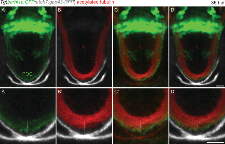
Barhl1a:GFP fibers co-localize with the neuronal projection marker acetylated Tubulin, a marker for the POC. (A–D) single optical sections (Z-stacks) through the anterior brain of a 35 hpf, Tg(barhl1a:GFP;atoh7:gap43-RFP) embryos immunostained with acetylated-Tubulin labelling the POC and its fibers (in red). Frontal view, dorsal is to the top. Barhl1a:GFP cells and their projections are shown in green whilst the optic nerves labelled by Atoh7:gap43-RFP are shown in gray. The optic nerves from each eye cross at the optic chiasm site, ventral and anterior to the POC, to then proceed along the optic tract. (A’–D’) higher magnification scan through the POC of the same embryo reveals partial overlap of the acetylated-Tubulin-labelled POC (highlighted by the white dashed line) and the Barhl1a:GFP fibers. Scale bars: (A–D) 50 µm, (A’–D’) 20 µm.
Acknowledgments
This image is the copyrighted work of the attributed author or publisher, and
ZFIN has permission only to display this image to its users.
Additional permissions should be obtained from the applicable author or publisher of the image.
Full text @ Sci. Rep.
Your Input Welcome
Thank you for submitting comments. Your input has been emailed to ZFIN curators who may contact you if
additional information is required.
Oops. Something went wrong. Please try again later.