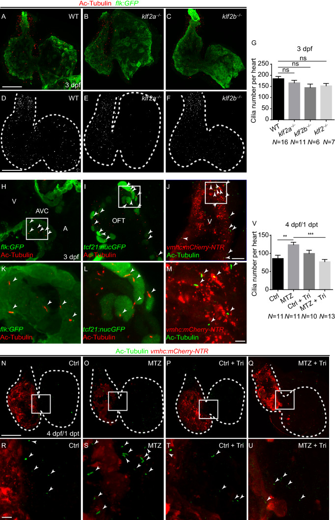
Endocardial primary cilia exist in the hearts at later stages. (A–F) Confocal stack projections of immunofluorescence showing cardiac cilia in wildtype and klf2 mutants at 3 dpf. Green, anti-GFP immunostaining; red, acetylated tubulin immunostaining. (D–F) greyscale of red channel only. (G) Quantification of cardiac cilia number in wildtype and klf2 mutants at 3 dpf (N = 16, 11, 6, 7 respectively). Mean + s.e.m. ANOVA analysis, ns, not significant. (H–M) Acetylated tubulin immunostaining showing primary cilia in endocardial cells (H, K), epicardial cells (I, L), and cardiomyocytes (J, M) at 3 dpf. (K–M) magnified white boxes in (H–J). (N–U) Confocal stack projections of immunofluorescence showing cardiac cilia in controlTricaine or BDM treatment or ablated Tg(vmhc:mCherry-NTR) hearts with or without Tricaine treatment at 1 dpt. (R–U) magnified white boxes in (N–Q). Red, mCherry fluorescence; green, acetylated tubulin immunostaining. (V) Quantification of cardiac cilia number in control or ablated hearts with or without Tricaine treatment at 1 dpt/4 dpf (N = 11, 11, 10, 13 respectively). Mean + s.e.m. ANOVA analysis, **P < 0.01, ***P < 0.001. Scale bars, (A–F, N–Q) 50 µm, (H–J) 10 µm, (K–M, R–U) 5 µm. dpf, days post fertilization, dpt, days post treatment. Dashed lines outline the hearts. Arrowheads point to cilia. A, atrium; AVC, atrioventricular canal; OFT, outflow tract; V, ventricle
This image is the copyrighted work of the attributed author or publisher, and
ZFIN has permission only to display this image to its users.
Additional permissions should be obtained from the applicable author or publisher of the image.
Full text @ Protein Cell
Your Input Welcome
Thank you for submitting comments. Your input has been emailed to ZFIN curators who may contact you if
additional information is required.
Oops. Something went wrong. Please try again later.