
|
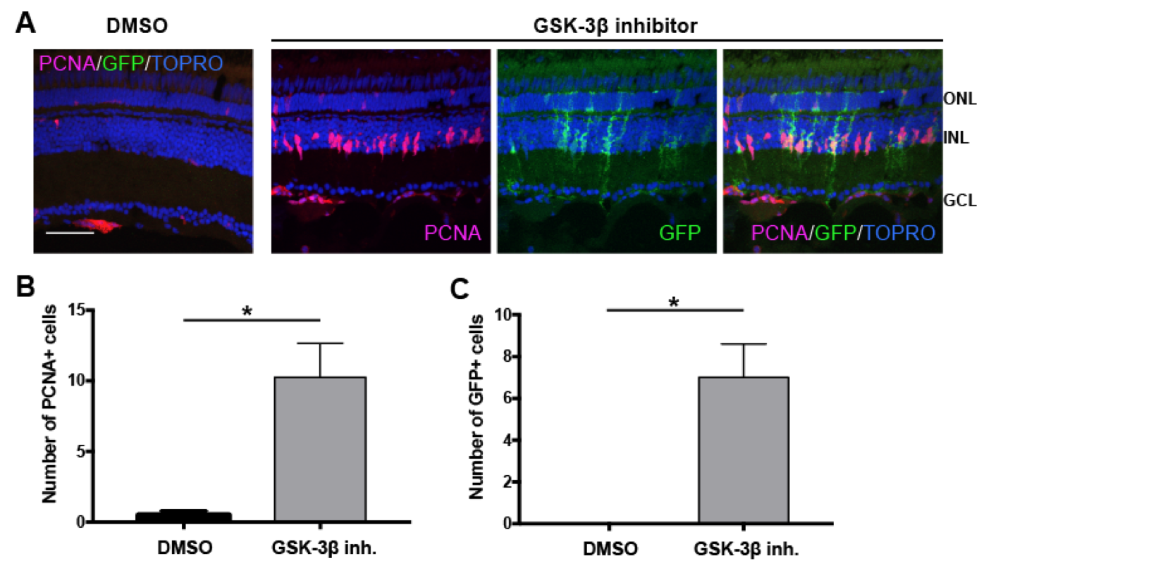
Fig. S4
β-catenin stabilization stimulates MG dedifferentiation and proliferation. Related to Figure 7. (A) Tg(1016tuba1a:gfp) adult fish were intravitreally injected with 1mM GSK-3β inhibitor (n=8) or control vehicle (DMSO) (n=4). Eyes were collected 51h post injection and sectioned retinas were immunostained using antibodies against GFP for dedifferentiated MG and PCNA for proliferating progenitors. Nuclei were counterstained with TOPRO (blue). (B) Quantification of PCNA+ proliferating progenitors and (C) GFP+ dedifferentiated MG in control vehicle and GSK-3β inhibitor injected retinas. Data represent mean +/- SEM, n= 4-8 fish; *, p<0.05 by two-tailed, Mann–Whitney U test. ONL, Outer nuclear layer; INL, inner nuclear layer; GCL, ganglion cell layer. Scale bar 50um.