
|
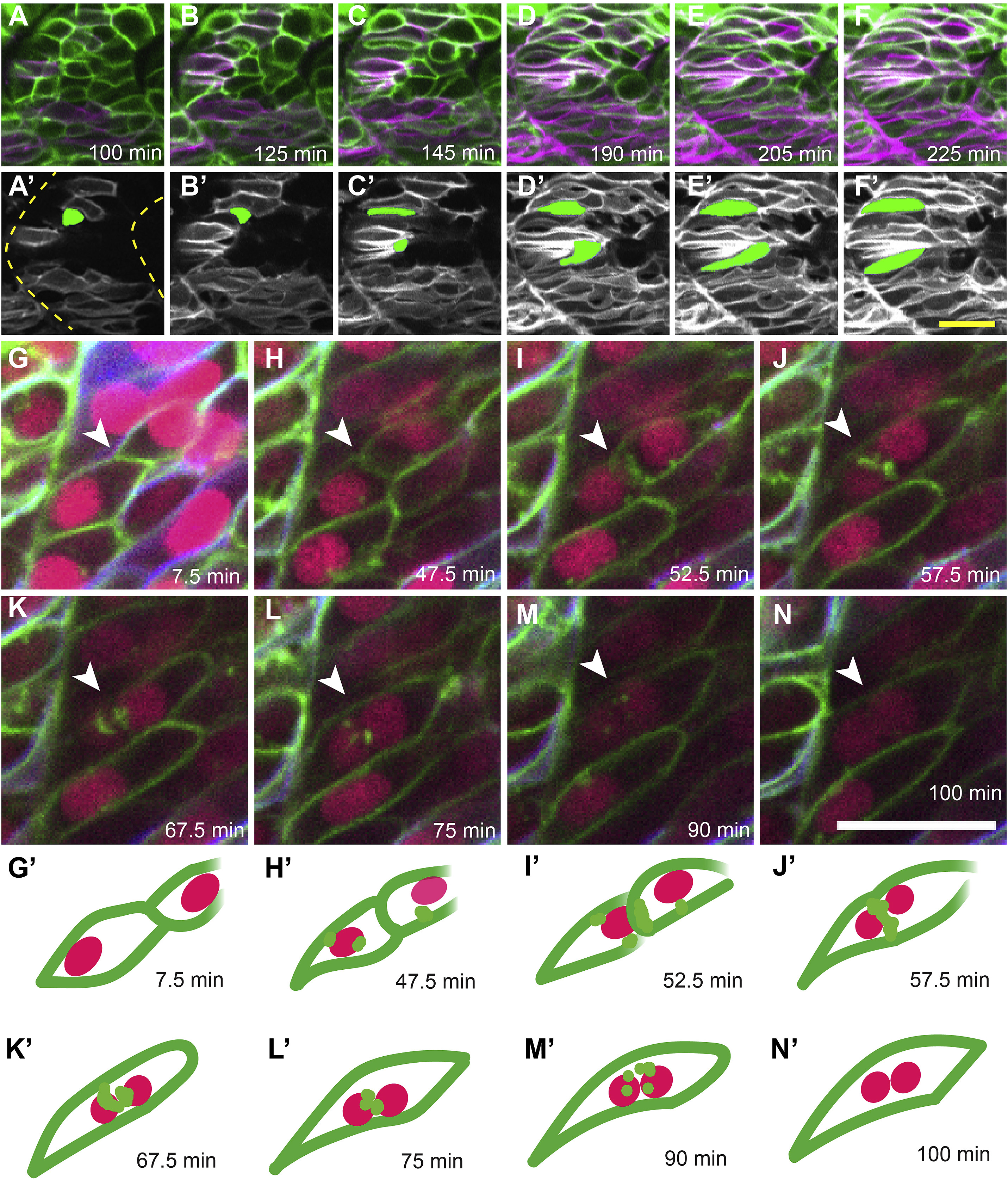
Fig. 1 Live imaging of slow muscle cell migration concurrent with fast muscle fusion events. (A–F) Frames from a time-lapse movie (Supplemental Movie 1) of a 19 hpf six1b:lyn-GFP (green); smyhc1:lyn-tdTomato (magenta) double transgenic embryo. (A′-F′) Pseudo-colored images show the slow muscle channel (white) from frames (A–F) overlaid with the shape of two fast muscle precursors (green) as they interact with slow muscle cells and extend anteriorly. Dotted outlines in A′ show somite boundaries (yellow). (G–N) Frames from a time-lapse movie of a 20 hpf six1b:lyn-GFP (green); smyhc1:lyn-tdTomato (blue) double transgenic embryo injected with mRNA encoding H2B-CFP (fuchsia). The movie (Supplemental Movie 2), which spans 92.5 min, was taken when fusion is actively occurring. (G′-N′) Illustrations depict a pair of fusing cells from Supplemental Movie 2 (G-N; white arrowhead). The time stamp of each frame is indicated. Scale bars in F’ (for A-F′) and N (for G-N) are 20 μm.
Reprinted from Developmental Biology, 462(1), Hromowyk, K.J., Talbot, J.C., Martin, B.L., Janssen, P.M.L., Amacher, S.L., Cell fusion is differentially regulated in zebrafish post-embryonic slow and fast muscle, 85-100, Copyright (2020) with permission from Elsevier. Full text @ Dev. Biol.