
|
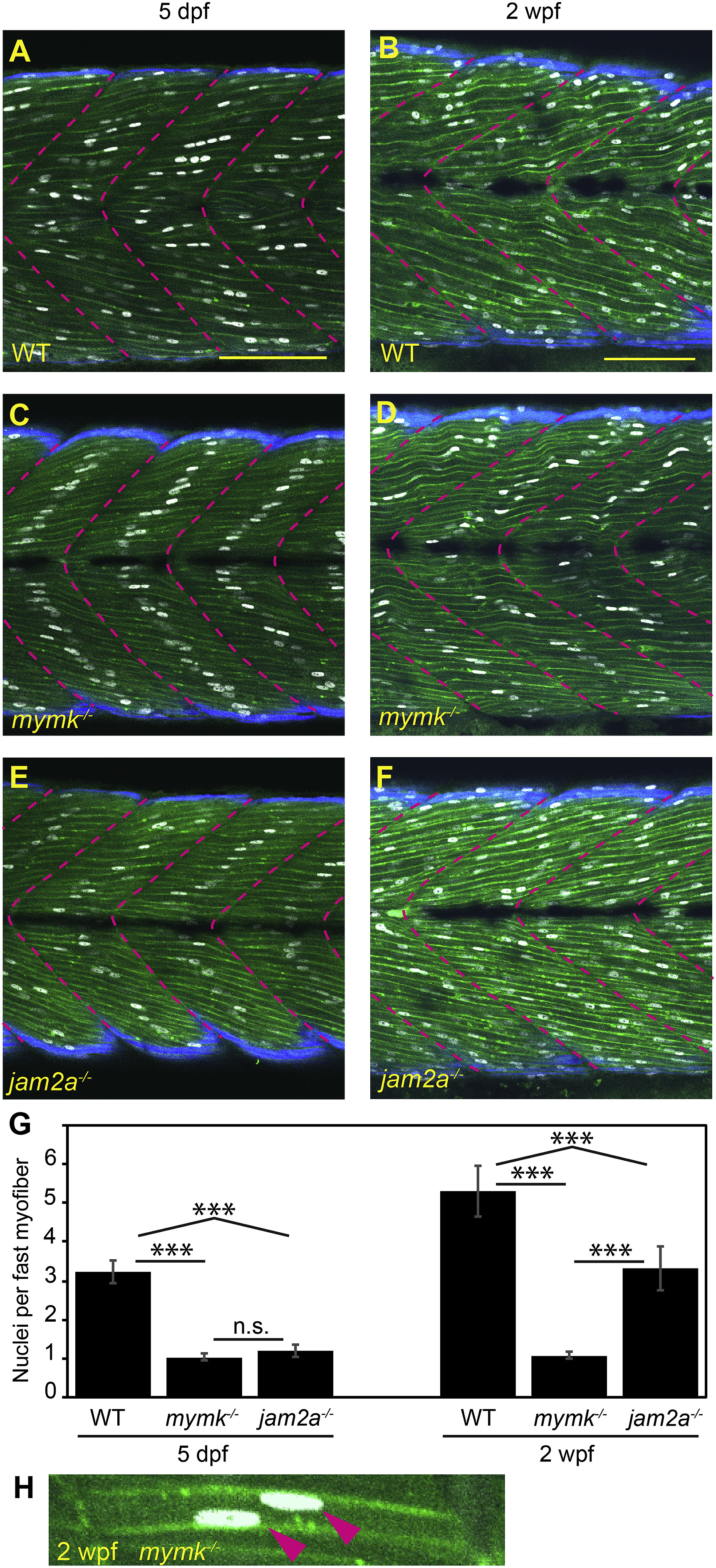
Fig. 3 jam2a is no longer required for multinucleation by 2 weeks post fertilization. (A–F) Sagittal confocal sections of 3MuscleGlow transgenic fish at 5 dpf (A, C, E) and 2 wpf (B, D, F). myog:H2B-mRFP (white) labels myonuclei, mylfpa:lyn-cyan (green) marks fast muscle cell membranes, and smyhc1:EGFP (aqua) marks slow muscle cells; myotomal boundaries are indicated by dotted lines (magenta). (G) Quantitative analysis reveals that mymk mutant myofibers are typically mononucleate at both stages. jam2a mutant myofibers, which are typically mononucleate at 5 dpf, are consistently multinucleate at 2 wpf. (H) An example of a rare mymk mutant fast myofiber with two nuclei (red arrows). Scale bars in A (for A, C, E) and B (for B, D, F) are 100 μm. Significance determined by ANOVA with Tukey-Kramer post-hoc analysis (p∗∗∗ < 0.001 compared to WT; n.s. indicates not significant).
Reprinted from Developmental Biology, 462(1), Hromowyk, K.J., Talbot, J.C., Martin, B.L., Janssen, P.M.L., Amacher, S.L., Cell fusion is differentially regulated in zebrafish post-embryonic slow and fast muscle, 85-100, Copyright (2020) with permission from Elsevier. Full text @ Dev. Biol.