
Fig. 8
- ID
- ZDB-IMAGE-200514-25
- Publication
- Saito et al., 2019 - Intraperitoneal dye injection method for visualizing the functioning lymphatic vascular system in zebrafish and medaka
- All Figures
- Figures for Saito et al., 2019
|
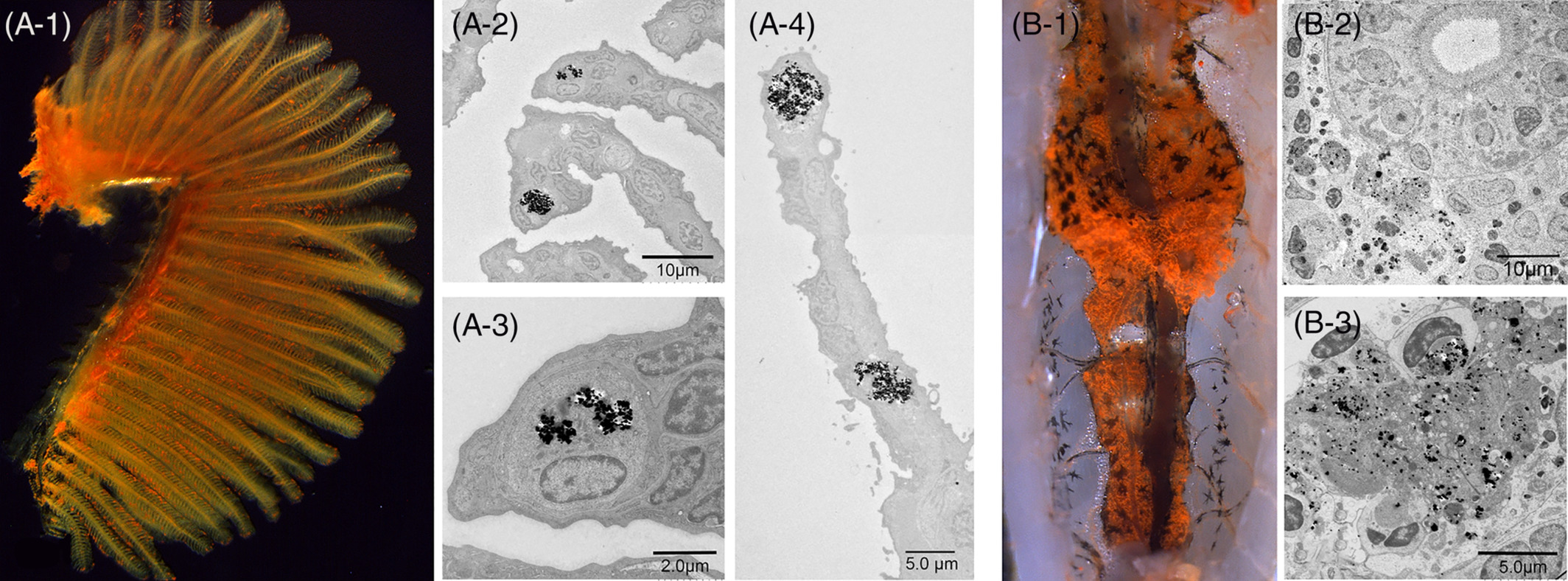
Fig. 8 Cinnabar dye in the gills (A‐1, left lateral view) and kidney (B‐1, ventral view) of adult zebrafish. The cinnabar dye predominantly accumulated in the gills and kidney, but the distribution pattern was easily distinguishable from that of lymphatic vessels by macroscopic visual examination. While the dye was distributed diffusely in the lymphatic ducts, it was identified as scattered red spots or granules in the gills (A‐1) and kidney (B‐1). As shown in the TEM images, monocyte/macrophage‐like cells took up the cinnabar particles in the secondary branchial lamella (A‐2,3,4) and in the blood‐forming tissue of the mesonephros (B‐2,3)