
|
Fig. 5
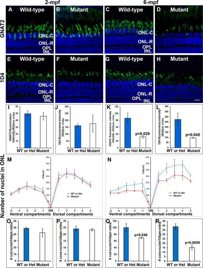
Loss of photoreceptors in pomgnt1 mutant retinas at 6-mpf but not 2-mpf. Retinal sections from 2-mpf and 6-mpf zebrafish were immunostained with GNAT2 antibody (green fluorescence, A–D) and 1D4 (green fluorescence, E–H). The sections were counter-stained with DAPI. To quantify photoreceptors, DAPI fluorescence confocal images of dorsal and ventral retinas from the optic nerve head to the ciliary margin were each divided into 5-equal compartments. Nuclei within the outer nuclear layer of each compartment were counted (M and N). GNAT2 and 1D4 immunoreactive intensities were measured (I–L). (A) Wild-type GNAT2 immunostaining at 2-mpf. (B) Homozygous pomgnt1sny7 mutant GNAT2 immunostaining at 2-mpf showing similar immunoreactivity compared to wild-type. (C) Wild-type GNAT2 immunostaining at 6-mpf. (D) Homozygous pomgnt1sny7 mutant GNAT2 immunostaining at 6-mpf showing reduced immunoreactivity compared to wild-type. (E) Wild-type 1D4 immunostaining at 2-mpf. (F) Homozygous pomgnt1sny7 mutant 1D4 immunostaining at 2-mpf showing similar immunoreactivity compared to the wild-type. (G) Wild-type 1D4 immunostaining at 6-mpf. (H) Homozygous pomgnt1sny7 mutant 1D4 immunostaining at 6-mpf showing reduced immunoreactivity compared to the wild-type. (I) GNAT2 immunofluorescence intensity quantification of wild-type and mutant fish at 2-mpf. There was no significant difference between wild-type and mutant retinas. (J) 1D4 immunofluorescence intensity quantification of wild-type and mutant fish at 2-mpf. There was no significant difference between wild-type and mutant retinas. (K and L) GNAT2 and 1D4 immunofluorescence intensity (artificial units, AU) in the mutant retina was reduced at 6-mpf. Student’s t-test. (M) Nuclei counting of outer nuclear layer at 2-mpf. There was no significant difference between wild-type and mutant retinas. (N) Nuclei counting of outer nuclear layer at 6-mpf. Nuclei within the outer nuclear layer in the mutant were reduced in number compared to wild-type. P = 0.00026; repeated measures ANOVA. (Oand P) 2-mpf nuclei count in the ONL-R and ONL-C retinal layers, respectively. There was no significant difference between wild-type and homozygous pomgnt1sny7 mutants. (Q and R) 6-mpf nuclei count in the ONL-R and ONL-C retinal layers, respectively. Note the significant reduction of nuclei in both ONL-R and ONL-C in homozygous pomgnt1sny7 mutants at 6-mpf. Together, these results indicated photoreceptor degeneration in mutant retina. Student’s t-test. Scale bar in H: 5 µm.