IMAGE

Figure 6

ID
ZDB-IMAGE-200509-19
Genes
Antibodies
Source
Figures for Furlan et al., 2020
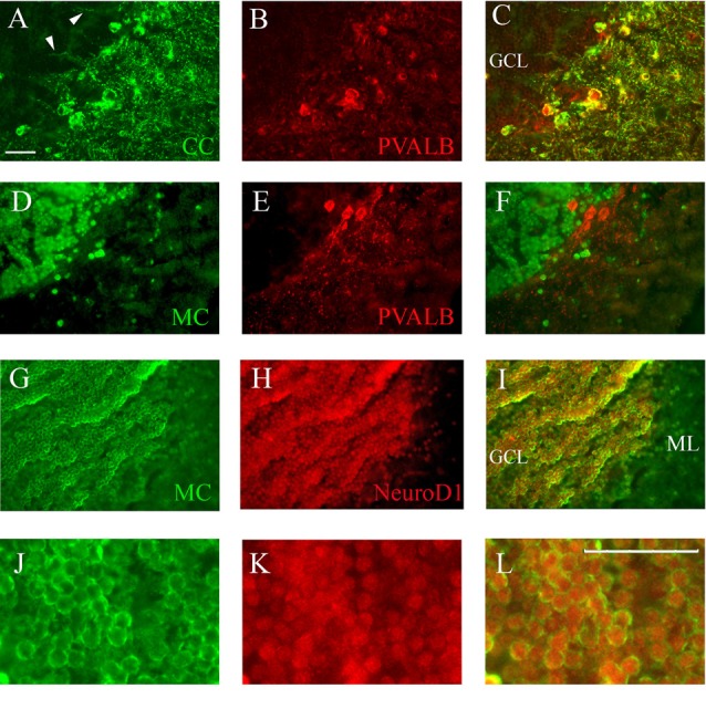
Image
Figure Caption

Figure 6

Co-staining of cerebellum with Casq and neuron-specific markers. (A–C) Epi-fluoresence signal obtained by double immunofluorescence with CC antibody (green) and anti-parvalbunin antibodies (red) in the cerebellum area. (C) A merge of the two images. Arrowheads indicate the axons of Purkinje cells. (D–F) Epi-fluoresence images obtained by double-staining with MC antibody (green) and anti-parvalbunin antibodies (red). (D) A merge of the two images. (G–I) Confocal images obtained by double-staining with MC antibody (green) and anti-NeuroD1 antibody (red). (J–L) Higher magnification of granule cells in the central area of the cerebellum double-labeled with MC (green) and anti-NeuroD1 antibody (red). Bar: 25 μm.

Figure Data
Acknowledgments
This image is the copyrighted work of the attributed author or publisher, and ZFIN has permission only to display this image to its users. Additional permissions should be obtained from the applicable author or publisher of the image. Full text @ Front. Neuroanat.