
Fig 6
- ID
- ZDB-IMAGE-200429-6
- Publication
- Arribat et al., 2020 - Spastin mutations impair coordination between lipid droplet dispersion and reticulum
- All Figures
- Figures for Arribat et al., 2020
|
Fig 6
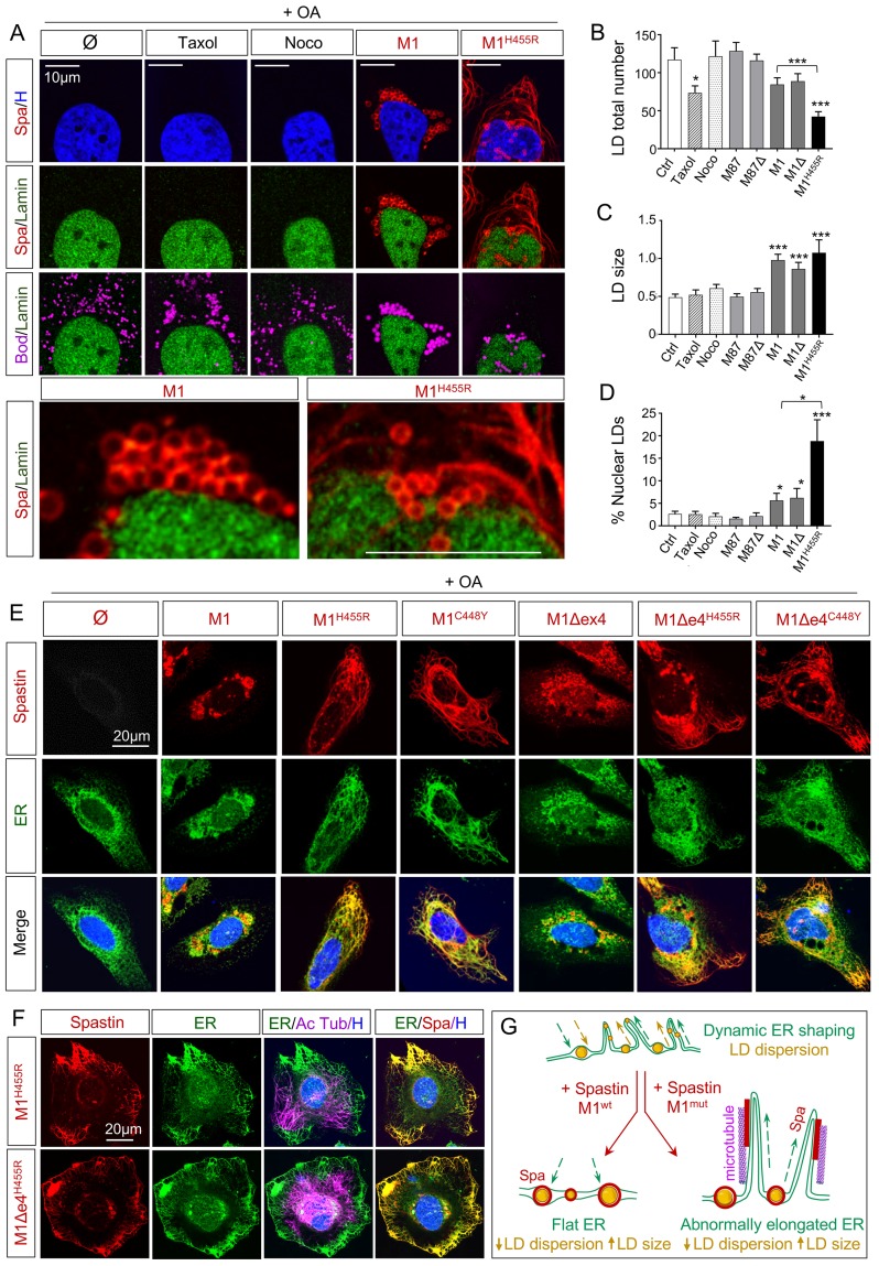
(A) Confocal microscopy images of HeLa cells overexpressing human Spastin M1 wild-type and mutant (H455R) treated with oleic acid (OA) for 18h. Nocodazole (Noco) and Taxol administration (12h) serve as control conditions. Cherry-tagged Spastin appears in red. Lamin A/C labeling corresponds to the nuclear compartment (green), Bodipy to LDs (magenta) and Hoechst to nucleus (blue). N = 4. (B-C) Quantification of LD number and size in HeLa cells treated with OA. n = 10 to 15 cells per condition. (D) Quantification of nuclear LDs in HeLa cells treated with OA. n = 10 to 15 cells per condition. (E-F) Confocal microscopy images of HeLa cells overexpressing human Spastin M1 isoforms treated with OA for 18h. Cherry-tagged Spastin appears in red. Calcineurin labeling corresponds to ER (green), acetylated Tubulin to microtubules (magenta) and Hoechst to nucleus (blue). (G) Schematic representation of ER shaping in presence of wild-type and mutant Spastin with subsequent impact on LD dispersion. In basal conditions, ER (green) follows a dynamic balance of elongation/retraction controlling the distribution of associated LDs (yellow) through the cell. Overexpression of wild-type Spastin M1 (red) triggers the retraction of ER leading to the accumulation of bigger LDs close to the nucleus. Mutated Spastin M1 (red) induces an abnormal elongation of ER along microtubules (magenta), resulting in a reduction of LD dispersion. Bars are mean ± SEM, *