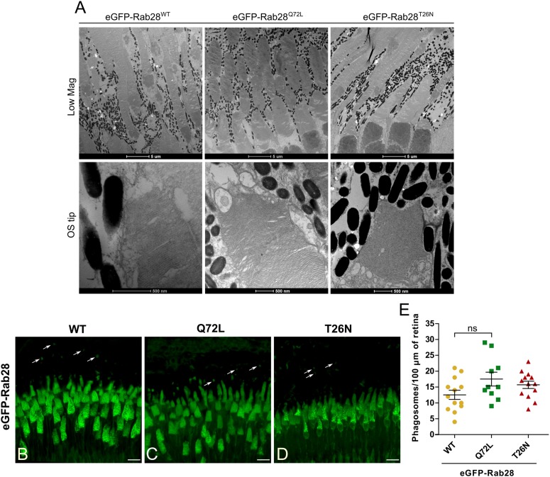
Rab28 transgenic zebrafish have normal retinal ultrastructure and normal outer segment shedding. (A) Representative TEM of 7 mpf eGFP-Rab28 transgenic zebrafish. Low magnification images show rows of several cone photoreceptors, while high magnification images show examples of OS tips. Low magnification scale bars 5 μm, high magnification scale bars 500 nm. (B–D) Confocal z-projections of 1 mpf retinas of zebrafish expressing eGFP-Rab28 (WT, Q72L, or T26N variants). Phagosomes, labeled with eGFP, are indicated by white arrows. Samples were collected 4 h after lights on. Scale bars 10 μm. (E) Scatter plots showing normalized phagosome density in the retinas eGFP-Rab28 transgenic zebrafish. Data are derived from cryosections in which eGFP was used to identify phagosomes. p-Value derived from one-way ANOVA. Error bars show SEM. Data are from 17, 17, and 13 retinal z-projections from at least three individuals for WT, Q72L, and T26N variants, respectively.
This image is the copyrighted work of the attributed author or publisher, and
ZFIN has permission only to display this image to its users.
Additional permissions should be obtained from the applicable author or publisher of the image.
Full text @ Front Cell Dev Biol
Your Input Welcome
Thank you for submitting comments. Your input has been emailed to ZFIN curators who may contact you if
additional information is required.
Oops. Something went wrong. Please try again later.