
|
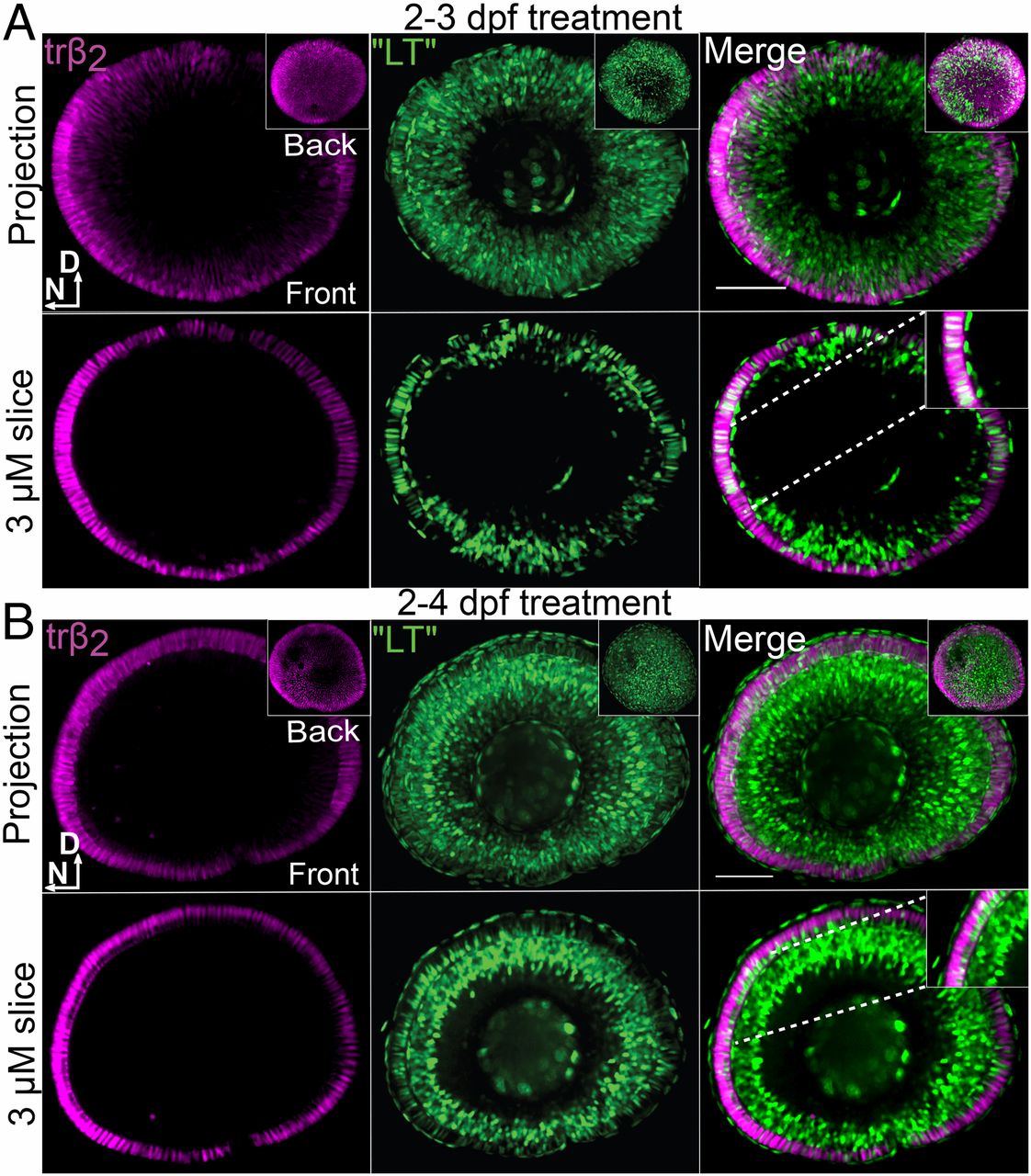
Fig. 4 TH “ligand trap” reporter transgenic and trβ2 reporter transgenic indicate accumulation of TH (T3) in lws+ (TRβ2+) cones after T3 treatment. All panels include visualization of T3 accumulation (ligand trap, GFP+), trβ2+ cones (tdTomato+), and merge (colabeled cells are white) of whole-mounted eyes visualized by confocal microscopy, from larvae treated with 100 nM T3 from 2 to 3 dpf (A) or 2 to 4 dpf (B). D, dorsal; N, nasal. Insets in Upper rows of A and B show views of the back of the eye; Insets in Lower rows of A and B show higher magnification of a single 3-µm z-slice to visualize coexpressing cones. (Scale bars, 50 μm.)