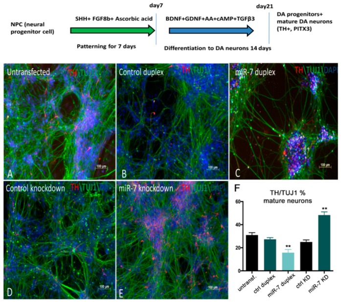
miR-7 negatively regulates the number of TH+ DA neurons derived from H9NPC cells. (A,B,C) Control H9NPC cells (A) showing TH staining at day 10 of DA neurogenesis (schematized in the top of the figure). BDNF: Brain Derived Neurotrophic Factor; GDNF: Glial cell Derived Neurotrophic Factor; cAMP: cyclic Adenosine MonoPhosphate; TGFβ3: Transforming Growth Factor beta 3. Decrease of TH+ cells (red) was observed when cells were transfected with miR-7 duplex (C), compared with the control duplex (B) and untransfected cells (A). (D,E) Increase of TH+ cells was observed upon knockdown of miR-7 (E), when compared with the control antisense. (F) Ratio of TH+/TUJ1 cells was quantified by manual counting of the neurons from images captured from 10 different areas on a random basis. Error bars represent average% ± SEM; the experiment was repeated thrice with n = 3 different biological replicates; (**) p < 0.01.
Acknowledgments
This image is the copyrighted work of the attributed author or publisher, and
ZFIN has permission only to display this image to its users.
Additional permissions should be obtained from the applicable author or publisher of the image.
Full text @ Cells
Your Input Welcome
Thank you for submitting comments. Your input has been emailed to ZFIN curators who may contact you if
additional information is required.
Oops. Something went wrong. Please try again later.